Fig. 7
- ID
- ZDB-FIG-150910-28
- Publication
- Toru? et al., 2015 - Endocytic Adaptor Protein Tollip Inhibits Canonical Wnt Signaling
- Other Figures
- All Figure Page
- Back to All Figure Page
|
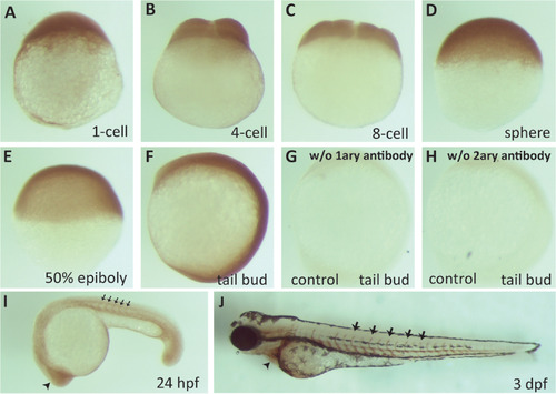
zTollip is broadly expressed during early zebrafish development. Immunocytochemistry analysis of zTollip protein localization in early embryonic development of zebrafish. Embryos fixed at various stages (indicated below the images) were incubated with primary antibodies against human Tollip, followed by secondary antibodies and chromogenic peroxidase-based detection. As controls for staining specificity, embryos at tail bud stage were processed omitting the primary antibody (G) or secondary antibody (H). zTollip expression is uniform from 1-cell stage until tail bud (A-F), but its expression increases in the head (arrowhead in I) and intersomitic regions (black arrows in I) at 24 hpf. At 3 dpf, zTollip is maintained in the intersomitic regions (arrows in J), but it is also expressed in the tissue surrounding the heart (arrowhead in J). Lateral views (A-H). Anterior to the left (I, J). |
| Antibody: | |
|---|---|
| Fish: | |
| Anatomical Terms: | |
| Stage Range: | 1-cell to Protruding-mouth |

