- Title
-
A Pak1/Erk Signaling Module Acts through Gata6 to Regulate Cardiovascular Development in Zebrafish
- Authors
- Kelly, M.L., Astsaturov, A., Rhodes, J., Chernoff, J.
- Source
- Full text @ Dev. Cell
|
Pak1 Is Required for Normal Development of Zebrafish (A) Transcripts for pak1 were detected by whole-mount in situ hybridization at the stages indicated. epib, epiboly. (B) Expression of pak1 at various stages in development by RT-PCR. (C) RT-PCR analysis of pak1 transcripts in morphants compared to WT and MM-injected embryos at 24 and 48 hpf. (D) Immunoblot analysis of Pak1 in morphants compared to WT at 24 and 48 hpf. (E) Representative images of the moderate (MO moderate) and severe (MO severe) pak1 knockdown phenotypes as compared to the WT and MM-injected embryos at 24 hpf and 48 hpf. (F) Quantification of pak1 knockdown phenotype at 24 hpf. p < 0.0001. (G) Morphology of embryos injected with the indicated MOs plus mRNA for WT of kinase-dead human Pak1 (hPak1), WT zebrafish Pak1 (zPak1), zPak2a, or zPak2b. (H) Quantitation of phenotypes at 24 hpf. p < 0.0001. EXPRESSION / LABELING:
|
|
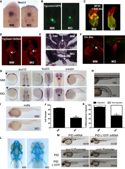
Pak1 Morphants Display Cardiac and Neural Crest Defects (A) Transcript expression of nkx2.5 shown by in situ hybridization at 20 somites. (B and C) Pak1 morphant heart does not loop at 48 hpf as shown by (B) Tg(cmlc2:GFP) immunofluorescence and (C) staining with MF20 and S46 antibodies. (D) Ventral images of 48 hpf Tg(Gata1:DsRed) show a cardiac outflow tract blockage with no blood flow throughout the head vasculature in the pak1 morphants (MO) compared to MM embryos (MM). Arrows point to cardiac outflow tract. (E) Transverse sections of the cardiac outflow tract in MO- and MM-injected embryos stained with H&E. Arrows point to the cardiac outflow tract. (F) Eln2 staining of WT and pak1 morphant embryos. Arrow indicates region of Eln positivity. Eln Abs, Eln2 antibodies. (G) In situ hybridization for sox10 and foxD3 at 16?18 somites (lateral and cranial views). Arrows point to cranial neural crest expression in lateral view. In situ hybridization for crestin at the 16- to 18-somite stage and the 20-somite stage. Note lack of migration of neural crest in cranial view (arrows) and down the tail in the lateral view. (H) Trunk images of pak1 morphants lacking melanophores at 2 dpf. (I) In situ hybridization for mitfa. (J) Quantification of melanophores at 2 dpf. Error bars indicate a significant difference with p < 0.0001. p < 0.0001. (K) Quantification of melanophore migration in pak1 morphants compared to MM-injected embryos. Error bars indicate a significant difference with p < 0.0001. (L) Cartilage staining (Alcian blue) at 6 dpf. (M) Top: WT embryos were injected with either PID (Pak inhibitor) or PID L107F (inactive Pak inhibitor) mRNA. Embryos are shown at 48 hpf. Bottom: WT embryos were injected with expression vectors encoding PID or PID L107F driven by the sox10 promoter or the cmlc promoter, as indicated. Images were taken at 48 hpf. EXPRESSION / LABELING:
PHENOTYPE:
|
|
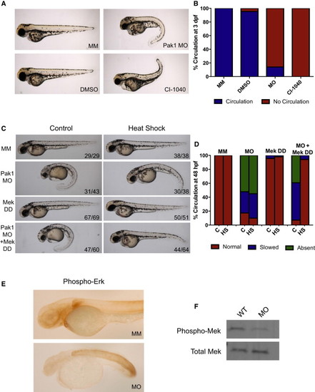
Pak1 Signals through the Erk Pathway in Heart Development (A) Comparison of chemical inhibition of Mek and pak1 morphants at 3 dpf. WT embryos were placed in egg water containing dimethyl sulfoxide (DMSO) or 0.5 ÁM CI-1040 at the one-cell stage. The water was changed every 24 hr with new drug. The embryos were analyzed for gross morphology and presence or absence of circulation. (B) Quantification of circulation seen with Mek inhibition at 48 hpf. (C) Representative images of 48 hpf pak1 morphants rescued by an active form of Mek (Mek DD). An inducible Mek1 DD expression plasmid was coinjected with the pak1 MO at the one-cell stage, followed by heat shock at 24 hpf as indicated. (D) Quantification of circulation seen with the addition of Mek DD. (E) Immunohistochemistry for phosphorylated Erk. (F) Immunoblot for phosphorylated Mek in whole embryos injected with either MM or pak1 MO. EXPRESSION / LABELING:
PHENOTYPE:
|
|
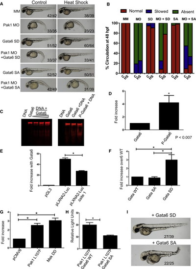
Active Gata6 Can Compensate for Loss of Pak1 (A) Representative images of pak1 morphants that have been rescued by the addition of an active form of Gata6 (Gata6 SD) but not by the inactive mutant (Gata6 SA). (B) Quantification of circulation in the Gata6 rescue. The indicated inducible Gata6 expression plasmids were coinjected with pak1 MO at the one-cell stage, followed by heat shock at 24 hpf as indicated. (C) Fluorescent EMSA showing dose-dependent binding of WT Gata6 to a GATA-containing DNA template (EMSA) as well as effects of Gata6 phosphorylation on DNA binding. (D) Quantitation of fluorescent EMSA showing that Gata6, when phosphorylated by ERK, is able to bind to target DNA more efficiently than unphosphorylated Gata6. (E) Luciferase assay showing transcriptional effects of Gata6 using WT or mutant GATA reporters. p < 0.05. (F) Luciferase assay comparing Gata6 SA and SD to WT. p < 0.05. (G) Luciferase assays comparing active Pak1 (Pak L107F) and MEK DD, respectively, to control. p < 0.05. (H) Luciferase assay comparing activated Pak1 plus either Gata6 WT or Gata6 SA. p < 0.05. (I) WT embryos were injected with a vector encoding PID driven by the sox10 promoter plus an expression vector encoding either Gata6 SD or SA. Representative images were taken at 48 hpf. PHENOTYPE:
|
|
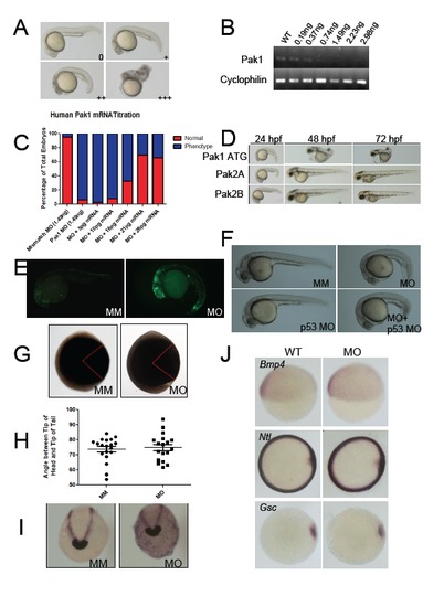
Characterization of pak1 morphant phenotype. (A) Representative images of embryos displaying mild, moderate, and severe phenotypes when injected with a titration of the pak1 morpholino. Table describing phenotypes and n numbers are located in Supplemental Table S1. (B) RT-PCR of embryos injected with a titration of pak1 morpholino. (C) Percentages of embryos displaying aberrant phenotypes when injected with the Pak1 morpholino in addition to varying amounts of human pak1 mRNA. (D) Representative images of embryos from 24-72 hpf injected with pak1 ATG MO, pak2a MO, or pak2b MO. Table describing phenotypes and n numbers are located in Supplemental Table S2. (E) Apoptotic cells were detected by a TUNEL assay on 24 hpf mismatch MO injected embryos or pak1 morphants. (F) Embryos co-injected with p53- MO and pak1 MO displayed a similar phenotype to pak1 morphants. (G) Representative images of 2 somite stage embryos injected with either a mismatch or pak1 MO, displaying angle between tip of head and tip of tail. (H) Quantification of gastrulation angles from panel A. No significant difference between control and pak1 morphants. (I) Embryos fixed at 10 hpf. In situ staining was performed with probes hgg1 and dlx3. (J) Staining at 6 hpf is shown for the ventral fate marker bmp4, mesodermal marker ntl, and the dorsalising factor gsc. Related to Figure 1. |
|
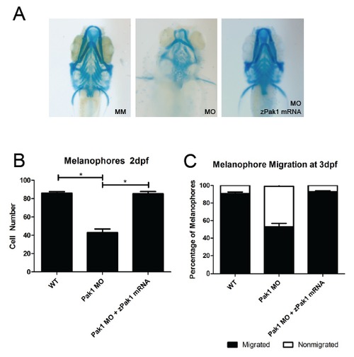
Zebrafish pak1 mRNA rescues the pak1 morphant cartilage and melanophore phenotype. Embryos were injected with 1.49 ng of the pak1 MO and 21 pg of zebrafish pak1 mRNA and subsequently analyzed for rescue of cartilage abnormalities through (A) Alcian blue staining, (B) the presence of melanophores, and (C) melanophore migration. Related to Figure 2. |
|
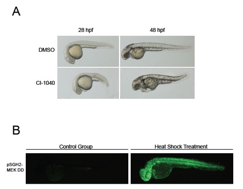
Mek signaling effects. (A) Effects of Mek inhibitor CI-1040 on embryonic development. CI-1040 was added at 0.5 ÁM to single cell embryos. (B) Ubiquitous transient expression following induction from the heat inducible vector, pSGH2. After injection into the embryo, pSGH2 can be activated by heat, inducing both GFP and the gene of interest, or MEK DD, in a ubiquitous manner. Related to Figure 3. |
Reprinted from Developmental Cell, 29, Kelly, M.L., Astsaturov, A., Rhodes, J., Chernoff, J., A Pak1/Erk Signaling Module Acts through Gata6 to Regulate Cardiovascular Development in Zebrafish, 350-9, Copyright (2014) with permission from Elsevier. Full text @ Dev. Cell


