- Title
-
Individual axons regulate the myelinating potential of single oligodendrocytes in vivo
- Authors
- Almeida, R.G., Czopka, T., Ffrench-Constant, C., and Lyons, D.A.
- Source
- Full text @ Development
|
Transgenic reporters reveal first axon myelinated in vivo in zebrafish CNS. (A) Lateral view of a stable Tg(mbp:EGFP-CAAX) zebrafish at 8 dpf. Myelinating glia of the CNS and PNS are labelled, as is the heart, which serves as a marker of transgenesis. (A′) Lateral view of the spinal cord (area indicated by box in A). Prominent myelinated tracts myelinated in the dorsal spinal cord and ventral spinal cord are apparent. (B) Lateral views of a stable Tg(mbp:EGFP-CAAX) zebrafish at 60 hpf show that the very first axon to be myelinated is the large Mauthner axon in the ventral spinal cord, which is first myelinated in the anterior spinal cord. (C) Lateral views of a stable Tg(mbp:EGFP-CAAX) zebrafish at 70 hpf. Myelination of the Mauthner axon has now commenced in the more posterior part of the spinal cord. At this stage, oligodendrocytes have started to myelinate axons in the dorsal spinal cord. Dorsal is up and anterior is to the left in all images. Scale bars: 500 μm in A; 20 μm in C. |
|
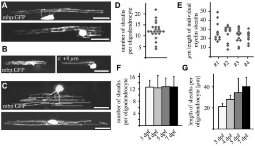
Single-cell analysis reveals morphological diversity of individual CNS oligodendrocytes. (A) Lateral views of single mbp:EGFP-expressing oligodendrocytes associating with one Mauthner axon (top) and both Mauthner axons (bottom). (B) Lateral view of a single mbp:EGFP-expressing oligodendrocyte associating with the large Mauthner axon (left) and an axon of much smaller caliber (right). (C) Lateral views of single oligodendrocytes associating with multiple axons in the dorsal spinal cord (top) and ventral spinal cord (bottom). (D) Myelin sheath number per oligodendrocyte (excluding those that myelinate the Mauthner axons) at 4 dpf. (E) Myelin sheath length in four sample oligodendrocytes (that do not associate with the Mauthner axon) at 4 dpf. (F) Average myelin sheath number per cell over time. This does not include oligodendrocytes that myelinate the Mauthner axon. (G) Average myelin sheath length per cell over time. This does not include oligodendrocytes that myelinate the Mauthner axon. Error bars represent s.d. Scale bars: 10 μm. |
|
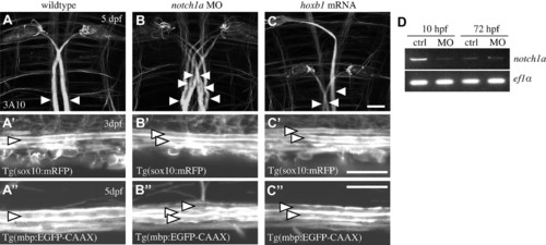
Supernumerary Mauthner axons are ensheathed by myelinating glia. (A-C) Ventral views of the hindbrains of wild-type (A), notch1a morphant (B) and hoxb1 mRNA-injected (C) larvae at 5 dpf labelled with the 3A10 antibody, which recognises neurofilament-associated proteins. Mauthner axons are indicated by arrowheads. Scale bar: 20 μm. (A′-C′) Analysis of Tg(sox10:mRFP) (A′-C′) and Tg(mbp:EGFP-CAAX) (A′-C′) shows that wild-type (A′,A′), notch1a morphant (B′,B′) and hoxb1-injected (C′,C′) Mauthner axons are all ensheathed and myelinated. Arrowheads indicate Mauthner axons. Scale bars: 20 μm. (D) RT-PCR analyses show that levels of notch1a mRNA are reduced in morpholino (MO)-injected animals relative to controls at 10 hpf but not at 72 hpf. |
|
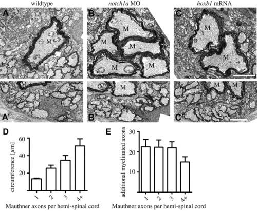
Supernumerary Mauthner axons are robustly myelinated. (A-C′) Transmission electron microscope images of transverse sections through the spinal cord of wild-type (A,A′), notch1a morphant (B,B′) and hoxb1 mRNA-injected (C,C′) zebrafish larvae at 9 dpf shows that all Mauthner axons are robustly myelinated (A-C) and that there is a similar number of axons myelinated in the ventral spinal cord despite the presence of supernumerary Mauthner (M) axons (A′-C′). Scale bars: 2 μm. (D) Total circumference of Mauthner axon(s) as a function of the number of Mauthner axons present per hemi-spinal cord. (E) Number of myelinated axons, excluding Mauthner axon(s), present in the ventral spinal cord as a function of the number of Mauthner axons present per hemi-spinal cord. Error bars represent s.d. |
|
Presence of supernumerary Mauthner axons does not affect oligodendrocyte number or distribution. (A-C) Lateral views of the spinal cords in Tg(mbp:EGFP)-expressing wild-type (A), notch1a morphant (B) and hoxb1 mRNA-injected (C) animals shows that oligodendrocyte number and distribution are not affected by the presence of supernumerary Mauthner axons. Arrows point to oligodendrocytes in the dorsal spinal cord with projections to the ventral spinal cord. Compare with Fig. 7B. Scale bar: 20 μm. (D). Total oligodendrocyte number per 425 μm length of tissue in the mid-trunk spinal cord of wild-type, notch1a morphant and hoxb1 mRNA-injected animals and, in grey, the number of oligodendrocytes specifically located in the dorsal domain of the spinal cord. Error bars represent s.d. |
|
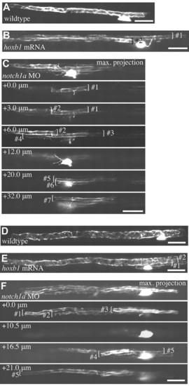
Individual oligodendrocytes myelinate multiple supernumerary Mauthner axons. (A-F) All images are of live single mbp:EGFP-expressing oligodendrocytes in the spinal cord of larvae at 5 dpf (A-C) and 9 dpf (D-F). (A,D) Wild-type oligodendrocytes associated with single Mauthner axons. (B,E) Oligodendrocytes in hoxb1 mRNA-injected animals with myelin sheaths on two Mauthner axons. (C,F) Oligodendrocytes in notch1a morphants that make seven (C) and five (F) myelin sheaths on supernumerary Mauthner axons. Maximum intensity projections and individual confocal z-sections are indicated for clarity. Individual myelin sheaths indicated by brackets. Scale bars: 20 μm. |
|
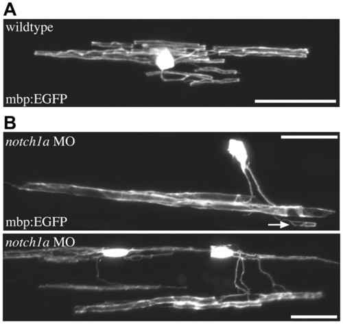
Individual oligodendrocytes readily myelinate supernumerary Mauthner axons and smaller caliber axons. (A) In wild type, oligodendrocytes in the dorsal spinal cord myelinate only small caliber axons. (B) Oligodendrocytes in the dorsal spinal cords of animals with supernumerary Mauthner axons can myelinate the large Mauthner axons in the ventral spinal cord in addition to smaller caliber axons (e.g. arrow). Scale bars: 20 μm. |
|
Tg(mbp:EGFP) labels myelinating glial in the CNS and PNS. (A) Lateral views of Tg(mbp:EGFP)-expressing zebrafish at 8 dpf. Anterior is to the left and dorsal up. (B) Schwann cells of the posterior lateral line, in the region indicated by the lower box in A. (C) High-magnification view of the spinal cord in the region indicated by the upper box in A. Note the Mauthner axon indicated by M. Scale bars: 500 μm in A; 10 μm in B,C. |


