- Title
-
Expression of ventral diencephalon-enriched genes in zebrafish
- Authors
- Li, S., Yin, M., Liu, S., Chen, Y., Yin, Y., Liu, T., and Zhou, J.
- Source
- Full text @ Dev. Dyn.
|
The expression profiles of a set of transcription factors in zebrafish larvae revealed by in situ hybridization. A: Id3 hybridization signals are detected from the two-cell stage. At later stages, Id3 expression is enriched in the diencephalon. An arrow indicates the polster, and arrowheads indicate the PT. Inserts are the dorsal view. B,C: Ebf2 and Ebf3 are highly expressed in the PT area of the diencephalon, MHB, OB, and HB. Arrowheads indicate the PT area of diencephalon. Dashed lines indicate the mid?hindbrain boundary. D: The expression of Klf7 is detected in the diencephalon, OB, DT, and HB. Inserts are lateral view of embryos with yolk sac and eyes removed. Arrowheads indicate the PT. E: Irx1 is specifically expressed in the midbrain and rhombomere 1 at 18 hours postfertilization (hpf). Dense signals were also detected at the Te, MHB, and kidney from 24 hpf. Arrows indicate the midbrain and the first rhombomere (r1). The insert is the dorsal view of the embryo. An arrowhead (in white) in the insert indicates the diencephalon. An asterisk indicates the kidney. DT, dorsal thalamus; HB, hindbrain; MHB, mid?hindbrain boundary; OB, olfactory bulb; PT, posterior tuberculum; Te, tectum. |
|
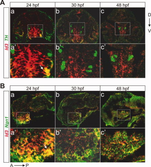
Id3 is expressed in the diencephalon and partially colocalized with Ngn1 or TH. A: Double labeled fluorescent in situ hybridization in coronal cryosections of larvae at 24, 30, and 48 hours postfertilization (hpf) reveals that Id3 (red) and TH (green) are co-expressed in a subset of cells in the diencephalon. a′?c′: The magnified views of the stippled boxes in a?c. B: Double labeling of Id3 and Ngn1 in sagittal cryosections of larvae at 24, 30, and 48 hpf. a?c, sagittal views of the embryos after double fluorescent in situ hybridization of Id3 (red) and Ngn1 (green). a′?c′, the magnified views of the stippled boxes in a?c. Arrows indicate the cells with colocalized hybridization signals. TH, tyrosine hydroxylase; D, dorsal; V, ventral; A, anterior; P, posterior. |
|
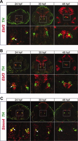
Ebf2, Ebf3, and Stmn4 mRNA are expressed in DA neurons in the PT area of the diencephalon. Larvae are subject to double-labeled in situ hybridization followed by coronal sections. A: Double-labeled in situ hybridization reveals that Ebf2 (red) and TH (green) are only colocalized in a subgroup of DA neurons at 24 hours postfertilization (hpf). Most Ebf2 signals surround TH signals. Lower panels are the enlarged views of the stippled boxes in the upper panels. B: Double-labeled in situ hybridization of Ebf3 and TH. They also show co-expression in a subgroup of DA neurons at 24 hpf. C: Double-labeled in situ hybridization of Stmn4 and TH. Colocalization is observed at 24, 30, and 48 hpf. Arrows in A, B, C indicate the cells with colocalized hybridization signals. DA, dopaminergic; TH, tyrosine hydroxylase; PT, posterior tuberculum. |
|
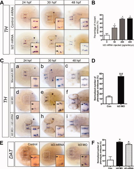
Altered Id3 expression levels result in a decreased number of TH+ cells at the PT area. A: TH in situ hybridization shows that the number of TH+ cells is decreased in Id3 mRNA-injected larvae. B: Quantification of the data shown in A. The graph shows the percentage of phenotypic embryos with reduced number of TH+ cells. C: TH in situ hybridization analysis in the larvae injected with control morpholino, Id3 morpholino, or Id3 morpholino with ?Id3 mRNA (Id3 mRNA lacking paired sequence with morpholino). The number of TH+ cells is also decreased in embryos injected with Id3 morpholino. Co-injection with ?Id3 mRNA can rescue these defects. D: Quantification of the data shown in C. Also the number of embryos with defects was quantified. E: In situ hybridization of DAT in manipulated embryos of 30 hours postfertilization (hpf). The signals of DAT are decreased in either Id3 mRNA- or MO-injected embryos. F: Quantitative data shown in E. The number of embryos with defects was quantified. Inserts in A, C, E are magnified views of the stippled boxes in white representing part of the diencephalon. Arrows indicate the PT with reduced TH+ cells. *P < 0.05. **P < 0.01. TH, tyrosine hydroxylase; MO, morpholino; PT, posterior tuberculum; DAT, dopamine transporter. |
|
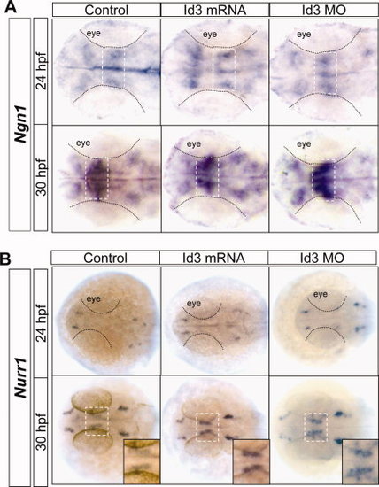
Id3 plays important roles in the maintenance and maturation of DA neurons. A: In situ hybridization reveals that Ngn1 expression in zebrafish larvae with either Id3 overexpression or Id3 knockdown is not markedly altered compared with control. Arrows indicated the diencephalon. B: In situ hybridization shows increased expression of Nurr1 at 30 hours postfertilization (hpf) in zebrafish larvae with either Id3 overexpression or knockdown compared with control. Inserts in A, B are magnified views of the stippled boxes in white indicating the diencephalon. MO, morpholino; DA, dopaminergic. |
|
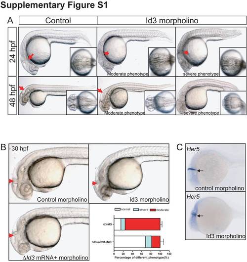
The phenotypes of Id3 morphants can be rescued by co-injection with Id3 mRNA. A: Phenotypes of Id3 morphants. Morphants show destroyed MHB at 24 hours postfertilization (hpf) and enlarged fourth ventricle at 48 hpf. The severe ones in the right panel show smaller a body. Arrowheads indicate the MHB, while arrows indicate the fourth ventricle. B: The defects caused by the Id3 morpholino can be rescued by ?Id3 mRNA. Arrows indicate the MHB. Quantitative analysis indicates that more than 70% of embryos injected with Id3 morpholino showed abnormal phenotype, whereas only approximately 30% of embryos co-injected with ?Id3 mRNA showed abnormal phenotypes. C: In situ hybridization of the MHB-specific gene Her5 indicates that the MHB is disturbed by Id3 morpholino. MHB, mid?hindbrain boundary. |


