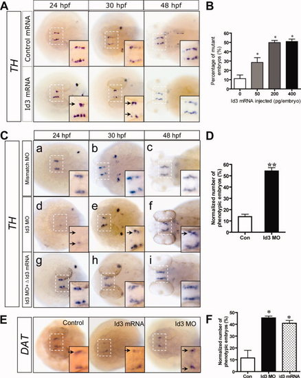
Altered Id3 expression levels result in a decreased number of TH+ cells at the PT area. A: TH in situ hybridization shows that the number of TH+ cells is decreased in Id3 mRNA-injected larvae. B: Quantification of the data shown in A. The graph shows the percentage of phenotypic embryos with reduced number of TH+ cells. C: TH in situ hybridization analysis in the larvae injected with control morpholino, Id3 morpholino, or Id3 morpholino with ?Id3 mRNA (Id3 mRNA lacking paired sequence with morpholino). The number of TH+ cells is also decreased in embryos injected with Id3 morpholino. Co-injection with ?Id3 mRNA can rescue these defects. D: Quantification of the data shown in C. Also the number of embryos with defects was quantified. E: In situ hybridization of DAT in manipulated embryos of 30 hours postfertilization (hpf). The signals of DAT are decreased in either Id3 mRNA- or MO-injected embryos. F: Quantitative data shown in E. The number of embryos with defects was quantified. Inserts in A, C, E are magnified views of the stippled boxes in white representing part of the diencephalon. Arrows indicate the PT with reduced TH+ cells. *P < 0.05. **P < 0.01. TH, tyrosine hydroxylase; MO, morpholino; PT, posterior tuberculum; DAT, dopamine transporter.
|
