- Title
-
TTLL3 Is a tubulin glycine ligase that regulates the assembly of cilia
- Authors
- Wloga, D., Webster, D.M., Rogowski, K., Bré, M.H., Levilliers, N., Jerka-Dziadosz, M., Janke, C., Dougan, S.T., and Gaertig, J.
- Source
- Full text @ Dev. Cell
|
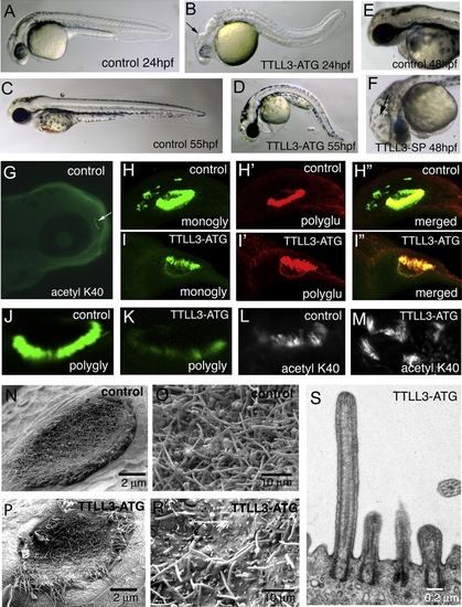
TTLL3 Is Required for Tubulin Glycylation and Cilia Assembly in Zebrafish (A–F) Images of live control and morphant embryos at 24 (A and B), 48 (E and F), and 55 hpf (C and D). Hydrocephaly is apparent in TTLL3-ATG morphant embryos, as indicated by the enlarged vesicles in the hindbrain (B and F, arrows). (G) The head of a WT embryo stained by 6-11 B1 antibodies at 72 hpf (the olfactory placode is marked with an arrow). (H–I″) In olfactory cilia, TTLL3-ATG MOs reduce the levels of tubulin monoglycylation detected by TAP952 mAb at 72 hpf (compare [H] and [I]), but not the levels of tubulin polyglutamylation revealed by polyE antibodies (H′ and I″). (J and K) The olfactory cilia stained with AXO49 anti-polyglycylated tubulin mAb in a control (J) and a TTLL3-ATG morphant (K) at 72 hpf. (L and M) Olfactory cilia stained with 6-11 B-1 mAb in a control (L) and a TTLL3-ATG morphant (M) at 72 hpf. (N–R) SEM images of the zebrafish olfactory placode at 72 hpf in control (N and O) and a TTLL3-ATG morphant (P and R); (O) and (R) are higher magnifications of areas of placodes shown in (N) and (P). (S) TEM image of a fragment of the olfactory placode in the TTLL3-ATG embryo. EXPRESSION / LABELING:
PHENOTYPE:
|
|
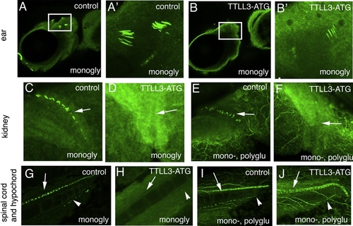
ttll3 MOs Reduce Tubulin Monoglycylation in Zebrafish Cilia (A–B′) Ear in a control (A and A′) and a morphant fish (B and B′) labeled with TAP952 mAb. (A′) and (B′) show higher magnifications (5x) of areas boxed in (A) and (B). (C–F) The pronephric duct area in control (C and E) and morphant fish (D and F) labeled with TAP952 anti-monoglycylated tubulin mAb (C and D) and GT335 anti-glutamylated tubulin mAb (E and F). Note lack of cilia staining in morphants in corresponding area (all indicated by arrows). (G–J) The spinal cord and hypochord area in control (G and I) and morphant fish (H and J) labeled with TAP952 (G and H) and GT335 mAb (I and J). Arrows indicate cilia in spinal cord, while arrowheads point to cilia in hypochord in control embryos (G and I). Note lack of cilia staining in morphants in corresponding regions, as indicated by arrows and arrowheads (H and J). EXPRESSION / LABELING:
|
|
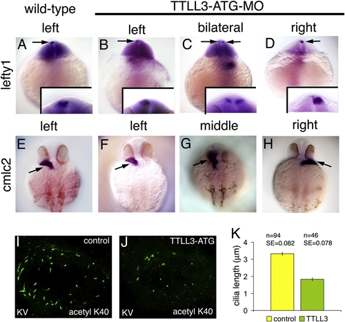
Depletion of ttll3 Expression Produces Defects in the LR Asymmetry in Zebrafish Ventral views of 22 hpf embryos stained for expression of lefty1 (A–D), or 33 hpf embryos stained for expression of cmlc2 (E–H). Insets in (A)–(D) depict 4x magnifications of the heads of embryos. In all cases, the left side of the embryo is to the left of the panel. lefty1 is normally expressed on the left side of the epiphysis (A). ttll3 morphant embryos express lefty1 on the left body side (B), or display bilateral (C) or right-sided expression (D). Arrows mark the epiphysis. In untreated embryos, cmlc2 (E) is expressed in the myocardium, which is normally located in the left lateral plate. The position of the heart field is unaltered in some TTLL3 morphants (F). Other morphants display midline (G) or right-side expression (H). Arrows in (E)–(H) mark the heart field. (I and J) Cilia in KV stained with 6-11 B-1 anti-acetylated K40 α-tubulin mAb in a control (I) and a TTLL3-ATG morphant (J) at 8-10 somite stage at 14 hpf. (K) A graph represents the average length of cilia in KV in control (n = 3) and TTLL3-ATG morphants (n = 3). Data are presented as means ± SEM. EXPRESSION / LABELING:
PHENOTYPE:
|
|
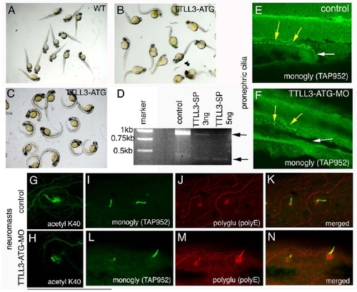
(A-C) A knockdown of TTLL3 expression in zebrafish changes fish morphology and affects cilia in multiple organs. Morphology of control embryos (A) and TTLL3-ATG morphants (B-C) at 48 hpf that were released manually from the chorion after 24 hpf (B) or at 72 hpf released manually from the chorion after 48 hpf (C). (D) Results of an RT-PCR assay for TTLL3 mRNA using total RNA from control and morphants injected with 3 or 5 ng of TTLL3-SP MO. (E and F). Depletion of TTLL3 reduces the TAP952 signal of tubulin monoglycylation in pronephron cilia; a control embryo (E) and a TTLL3-ATG morphant (F). (G-N) TTLL3 depletion shortens cilia and reduces the TAP952-positive tubulin monoglycylation. G and H are images of neuromasts stained with 6-11 B-1 anti-acetylated α- tubulin antibodies in control (G) and a TTLL3-ATG morphant (H). (I-N) Double labeling of control embryos (I-K) and TTLL3-ATG morphants (L-N) with TAP952 (I and L) and polyE anti-polyglutamylation antibodies (J and M). (K and N) Merged images. Note a decrease in the level of tubulin monoglycylation and increase in the level of tubulin polyglutamylation in morphants. |
|
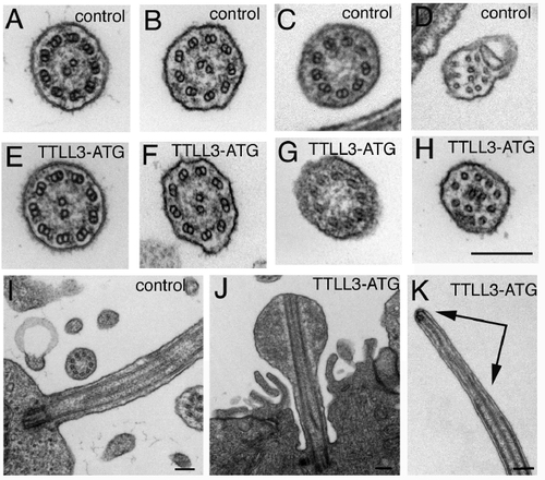
TEM study of cilia in the olfactory placode in a control (A-D, I) and in a morphant (E-H, J-K). Note that in a control placode, there are axoneme cross-sections that have both inner and outer (A) or only inner (B) dynein arms and some cross-sections lack the central pair (C). The distal ends of control cilia were contain the A-subfiber extensions (D). Ultrastructure of the remaining few morphant cilia was normal except that longitudinal sections show that some cilia have swollen tips (J). |
|
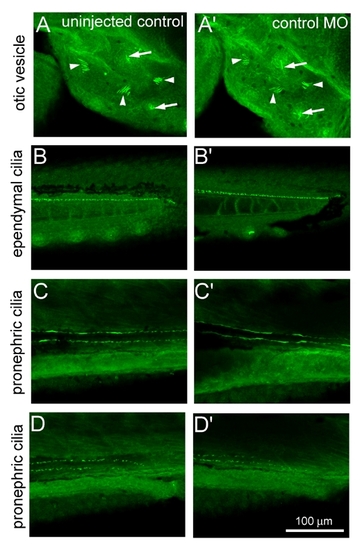
Injection of Random Sequence Morpholinos into Zebrafish Embryos Does Not Affect the Levels of Monoglycylation Detected by TAP952 Uninjected embryos (A-D) and embryos injected with 1ng of random sequence morpholinos (A′- D′), were fixed at 72 hpf , stained with TAP952 antibodies and the levels of tubulin monoglycylation were analyzed by immunofluoresce in cilia in the otic vesicle (arrowheads) and neuromasts (arrows) (A-A′), spinal cord (B-B′) and in middle (C-C′) and distal (D-D′) part of the pronephric duct.. Scale 100 μm. |
Reprinted from Developmental Cell, 16(6), Wloga, D., Webster, D.M., Rogowski, K., Bré, M.H., Levilliers, N., Jerka-Dziadosz, M., Janke, C., Dougan, S.T., and Gaertig, J., TTLL3 Is a tubulin glycine ligase that regulates the assembly of cilia, 867-876, Copyright (2009) with permission from Elsevier. Full text @ Dev. Cell

