- Title
-
Genetic dissection of neural circuits by Tol2 transposon-mediated Gal4 gene and enhancer trapping in zebrafish
- Authors
- Asakawa, K., Suster, M.L., Mizusawa, K., Nagayoshi, S., Kotani, T., Urasaki, A., Kishimoto, Y., Hibi, M., and Kawakami, K.
- Source
- Full text @ Proc. Natl. Acad. Sci. USA
|
The gene trap and enhancer trap constructs and the UAS reporter system. The Tol2 vector sequences are shown as thick black lines. (A) The structures of T2KhspGGFF, T2KhspGGFF, and T2KSAGFF. (B) The UAS:GFP reporter fish carries a single-copy insertion of T2KUASGFP within a gene encoding a homolog of Nedd4-binding protein 1. Blue boxes indicate exons. (C) The UAS:RFP reporter fish carries a single-copy insertion of T2ZUASRFP within a gene encoding a solute carrier protein homolog. Blue boxes indicate exons. (D and E) The hspGGFF1B embryos before (D) and after (E) heat shock. (F and G) The hspGGFF1B;UAS:GFP embryos before (F) and after (G) heat shock. (H and I) The hspGGFF1B;UAS:RFP embryos before (H) and after (I) heat shock. EXPRESSION / LABELING:
Constructs:
Et(T2KSAGFFLF)
,
Et(hsp70l:GFP-GAL4FF)
,
Gt(GAL4FF)
,
Tg(5xUAS:RFP)
,
Tg(5xUAS:EGFP)
,
Et(hsp70l:GAL4FF)
|
|
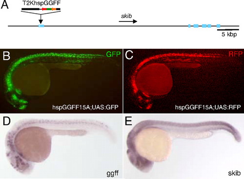
GFP and RFP expression in the hspGGFF15A enhancer trap line. (A) The structure of the hspGGFF15A insertion. T2KhspGGFF is integrated in the first exon of the skib gene. (B) GFP expression in the hspGGFF15A;UAS:GFP embryo at 24 hpf. (C) RFP expression in the hspGGFF15A;UAS:RFP embryo at 24 hpf. (D) Whole-mount in situ hybridization of the hspGGFF15A embryo at 24 hpf using the GGFF probe. (E) Whole-mount in situ hybridization of a wild-type embryo at 24 hpf and the skib probe. EXPRESSION / LABELING:
|
|
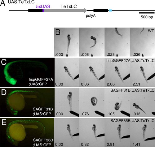
Abnormal touch response phenotypes in the UAS:TeTxLC double transgenic embryos. (A) The UAS:TeTxLC effector fish carries a single-copy insertion of T2MUASTeTxLC in the myosin heavy chain gene. A blue box indicates an exon. (B) The touch response behavior of a wild-type embryo at 48 hpf. (C) GFP expression in the hspGGFF27A;UAS:GFP embryo at 24 hpf and the touch response behavior of the hspGGFF27A;UAS:TeTxLC embryo at 48 hpf. (D) GFP expression in the SAGFF31B;UAS:GFP embryo at 24 hpf and the touch response behavior of the SAGFF31B;UAS:TeTxLC embryo at 48 hpf. (E) GFP expression in the SAGFF36B;UAS:GFP embryo at 24 hpf and the touch response behavior of the SAGFF36B;UAS:TeTxLC embryo at 48 hpf. |
|
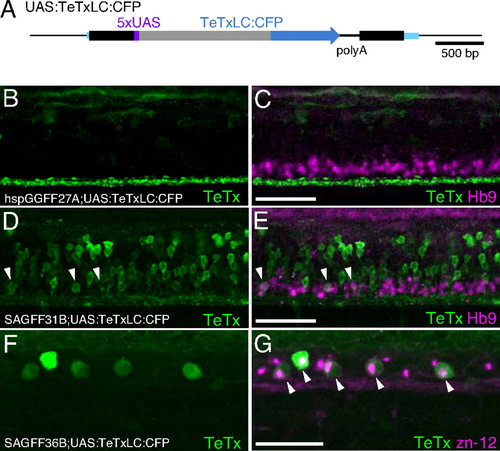
Expression of TeTxLC:CFP in double transgenic embryos. (A) The UAS:TeTxLC:CFP fish carries a single-copy insertion of T2SUASTeTxLCCFP within the CSPP1 gene. Blue boxes indicate exons. (B-G) Lateral views of the trunk of double transgenic embryos immunostained with the anti-GFP antibody (green) and the anti-Hb9 (B-E) or the zn-12 (F and G) antibody (red). Arrowheads indicate costaining with anti-GFP and anti-Hb9 (E) or anti-GFP and zn-12 (G). Anterior is to the left, and dorsal is to the top. (Scale bars: 50 Ám.) (B and C) The hspGGFF27A;UAS:TeTxLC:CFP embryo at 48 hpf. The anti-GFP antibody detects the TeTxLC:CFP fusion protein but does not detect the GGFF protein in this condition (data not shown). Axons of descending hindbrain interneurons were strongly stained (green). The anti-Hb9 antibody detected spinal motor neurons (red). (D and E) The SAGFF31B;UAS:TeTxLC:CFP embryo at 48 hpf. The anti-GFP antibody detected spinal interneurons and motor neurons (green). (F and G) The SAGFF36B;UAS:TeTxLC:CFP embryo at 30 hpf. Both the anti-GFP and zn-12 antibodies detected Rohon-Beard neurons (green and red).
Construct:
Tg(5xUAS:tetxlc-CFP)
|
|
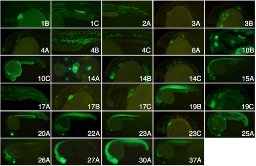
The GFP expression patterns in the hspGGFF;UAS:GFP and SAGFF;UAS:GFP embryos at 24 hpf. Offspring from crosses between these gene trap and enhancer trap fish lines and the UAS:TeTxLC effector line showed abnormal touch response behaviors. EXPRESSION / LABELING:
|
|
The TeTxLC:CFP expression in the hspGGFF27A;UAS:TeTxLC:CFP embryo at 24 hpf. Shown is a lateral view of the spinal cord of a 24-hpf hspGGFF27A;UAS:TeTxLC:CFP embryo immunostained with the anti-GFP antibody (green). The anti-GFP antibody detects the TeTxLC:CFP fusion protein but does not detect the GGFF protein in this condition (data not shown). Axons of descending hindbrain interneurons (arrowhead) and cell bodies of spinal neurons were detected. Anterior is to the left, and dorsal is to the top. (Scale bar: 50 mm.) EXPRESSION / LABELING:
|

Unillustrated author statements |


