- Title
-
Identification of new netrin family members in zebrafish: Developmental expression of netrin2 and netrin4
- Authors
- Park, K.W., Urness, L.D., Senchuk, M.M., Colvin, C.J., Wythe, J.D., Chien, C.B., and Li, D.Y.
- Source
- Full text @ Dev. Dyn.
|
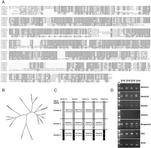
1. ClustalW amino acid alignment of Netrin sequences and reverse transcriptase-polymerase chain reaction (RT-PCR) expression analysis of Netrin and Netrin receptor genes. A: Alignment of chick Netrin2 and mouse Netrin4 with zebrafish Netrin2 and Netrin4. Dark and pale gray boxes represent, respectively, identical and similar amino acids. zf, zebrafish; m, mouse; c, chick. B: Phylogenetic analysis of predicted zebrafish Netrin proteins based on ClustalW alignment using the Biology Workbench site (http://workbench.sdsc.edu). zf, zebrafish; h, human; m, mouse; c, chick; dr, Drosophila. Zebrafish Netrin2 is most closely related to chick Netrin2, whereas zebrafish Netrin4 is closely related to human and mouse Netrin4. The GPI-linked proteins Netrin-G1 and -G2 are included for comparison. C: Zebrafish Netrin2 and Netrin4 contain all the domains (domains VI, V, and C) described in other Netrin family members. Numbers indicate identity, and parentheses represent similarities between two given proteins. Netrin2 is highly similar to Netrin1a and 1b, while Netrin4 is the most divergent member of the Netrin family. D: RT-PCR analyses were performed to detect netrin transcripts in total RNA collected from 24, 36, 48, and 72 hpf embryos. Actin amplification served as a loading control, and mock amplifications (-, no RT) confirmed that bands represent amplification of RNA. Netrin2 is expressed from 24 hpf through 72 hpf, whereas netrin4 is not detected at any of these stages (25 cycles). Netrin1a, netrin1b, dcc, and neogenin1 are expressed at all stages. |
|
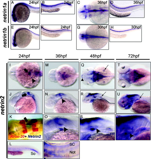
Netrin mRNA expression in zebrafish embryos. Netrin1a, netrin1b, and netrin2 expression domains were analyzed by RNA in situ hybridization during the first 3 days of embryonic development. A-H: Lateral (A,B,D-F,H) and dorsal (C,G) views of netrin1a (A-D) and netrin1b (E-H). A,B: netrin1a is expressed in the brain, spinal cord, and somites at 24 hpf. C,D: By 36 hpf, somite expression has diminished and expression is visible in the optic nerve. E,F: netrin1b is detected in the ventral brain, the floor plate of the spinal cord, and the hypochord at 24 hpf. G,H: Hypochord expression diminishes by 30 hpf. I-V: Dorsal (I,K,M,Q,T) and lateral (J,L,N-P,R,S,U,V) views of netrin2 expression at stages indicated. I,J: netrin2 is expressed in the fourth rhombomere (arrow) and in the otic vesicle (arrowhead) at 24 hpf. K: krox20 two-color in situ hybridizations confirm rhombomere 4 localization of netrin2 (arrow). L: netrin2 is expressed in the ventral somites (So) at 24 hours postfertilization (hpf). M,N: At 36 hpf, netrin2 expression extends to the dorsal and ventral margins of the otic vesicle (arrowhead) and in the hindbrain region. O: Enlarged view of the hindbrain and otic vesicle (arrowhead). P: netrin2 is also detected in notochord (Not) and spinal cord (SC) after prolonged colorimetric development. Q,R: At 48 hpf, netrin2 is expressed in bilateral clusters in the telencephalon (small arrowhead), around the medial and caudal margins of the tectum, in the cerebellum, and along the dorsal hindbrain, and in the ventromedial margin of the otic vesicles. It is also expressed in the pharyngeal arches and pectoral fins (small arrows). In contrast to netrin1a and 1b, expression in the brain is primarily constrained to the dorsal aspect. S: Higher magnification views of the hindbrain and otic vesicle; arrowhead denotes the strong expression in the ventral margin of the otic epithelium. T,U: By 72 hpf, expression in the pharyngeal arches is robust, and expression in the dorsal aspect of the otic epithelium and pectoral fins (small arrow) has strengthened. V: Higher magnification view of the otic vesicle; the arrowhead denotes the strong expression in the developing inner ear. EXPRESSION / LABELING:
|
|
EXPRESSION / LABELING:
|


