- Title
-
Phox2b function in the enteric nervous system is conserved in zebrafish and is sox10-dependent
- Authors
- Elworthy, S., Pinto, J.P., Pettifer, A., Cancela, M.L., and Kelsh, R.N.
- Source
- Full text @ Mech. Dev.
|
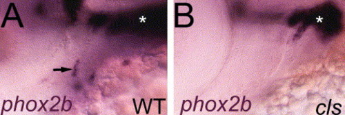
Expression of phox2b in the zebrafish branchial arches. Lateral views of whole-mount phox2b mRNA in situ hybridization in 60 hpf embryos shows strong expression in epibranchial ganglia (asterisk) in both wild-types (A) and sox10 mutant embryos (B). phox2b expression was also seen in isolated cells associated with the more ventral branchial arches (arrow, A), but these were absent in sox10 mutants (B). EXPRESSION / LABELING:
|
|
Late embryonic development of the zebrafish enteric nervous system. Lateral views of whole-mount (A, B, F?K) and transverse sections (C?E) of mRNA in situ hybridizations (A?E) or immunofluorescent detection (I?K) and corresponding DIC images (F?H) of markers of ENS progenitors in hindgut region. From 60 hpf, sox10 (A, C), phox2b (B, D) and ret (E) expression is prominent in two chains of ENS progenitors migrating posteriorly along the gut primordium (g). Note that sox10 expression at this stage is seen in cells right up to the anus (a), whereas phox2b (and ret, data not shown) is not expressed this far posteriorly (A, B). In contrast, expression of Hu antigen is not seen in the hindgut at 75 hpf (I), but is seen in increasing numbers of cells at 100 (J) and 120 hpf (K). EXPRESSION / LABELING:
|
|
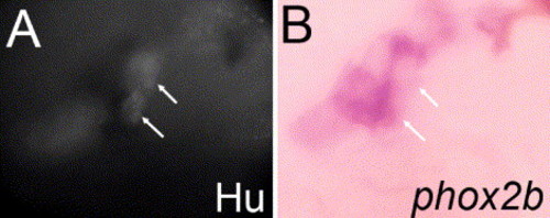
Expression of phox2b in enteric neurons and non-neuronal cells. (A, B) Dorsolateral views of ventral trunk to show ENS progenitors in 55 hpf embryos. Combined immunofluorescent detection of Hu antigen (A) and mRNA in situ hybridization for phox2b (purple, B) reveals double-labeled early differentiating neurons (arrows) containing both markers. Other cells express only phox2b and are presumably undifferentiated progenitors. EXPRESSION / LABELING:
|
|
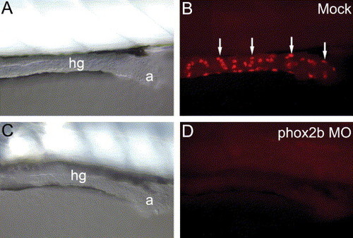
Injection of phox2b morpholinos generates partial phenocopies of the sox10 enteric neuron phenotype. Lateral views of posterior hindgut of 5 dpf wild-type embryos injected with 2 ng dose of phox2b.a (C, D) morpholino to show severe reduction in numbers of Hu-positive enteric neurons (arrows) compared with uninjected controls (A, B). DIC images of same embryos confirm their normal gross morphology (A, C). hg, hindgut; a, anus. PHENOTYPE:
|
|
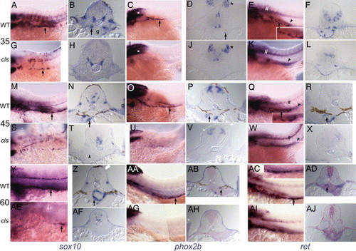
sox10 mutants show severe and early defects in enteric nervous system development. Lateral views (A, C, E, G, I, K, M, O, Q, S, U, W, Y, AA, AC, AE, AG, AI) or transverse sections (B, D, F, H, J, L, N, P, R, T, V, X, Z, AB, AD, AF, AH, AJ) of whole-mount in situ hybridizations of anterior trunk at 35 (A?L), 45 (M?X) and 60 hpf (Y?AJ) for enteric nervous system markers sox10 (A, B, G, H, M, N, S, T, Y, Z, AE, AF), phox2b (C, D, I, J, O, P, U, V, AA, AB, AG, AH) or ret (E, F, K, L, Q, R, W, X, AC, AD, AI, AJ) in wild-type (A?F, M?R, Y?AD) and sox10 (cls) mutant (G?L, S?X, AE?AJ) embryos. At all stages, wild-type ENS progenitors express all three genes (arrows), whereas sox10 mutants show occasional sox10-positive cells in this location, but do not express phox2b nor ret. As an internal positive control, note that expression of phox2b in the hindbrain (*, C, D, I, J) and ret in the ventral neural tube (#, E, K) is not affected. Note too that in wild-type embryos ret is expressed in both a chain of ENS progenitor cells (arrow and inset in E, Q and AC) and more medially in the tightly packed cells of the forming pronephros (partially out of focus in this focal plane; arrowhead, E, Q); the former is absent in sox10 mutants, whereas the latter is unaffected (K, W, AI). ENS progenitor marker expression extends more posteriorly along the gut with increasing age (e.g. compare C, O, AA). sox10 expression consistently extends more posteriorly than the other two markers at equivalent stages (e.g. compare A, C, E). Transverse sections reveal changes in progenitor distribution around gut primordium with age. ENS progenitors at 35 hpf initially lie in contact with gut primordium (arrow, B), forming two longitudinal columns migrating along the gut, but have spread around the developing gut by 60 hpf (Z). Note the identical positioning of sox10, phox2b and ret positive progenitors with respect to the developing gut (e.g. N, P, R). ENS progenitors are not usually seen in sections of cls mutant embryos (H, J, L, T, V, X, AF, AH, AJ), although expression in CNS remains. Interestingly, sox10 and phox2b expression is prominent and overlapping in the pectoral fin mesenchyme (p) of wild-types (N, P), whereas phox2b appears to be strongly reduced in cls mutants (T, V). NB All whole-mount embryos treated with PTU to inhibit melanin synthesis. g, gut primordium. |
|
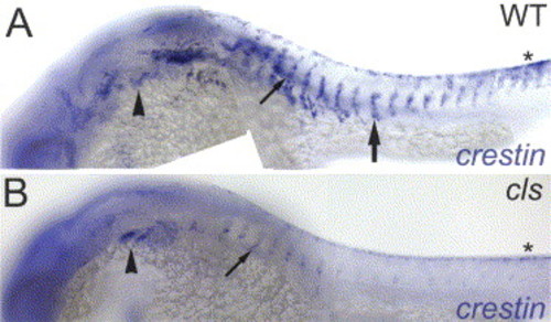
Widespread defects in crestin expression in sox10 mutants extend to premigratory stages of neural crest development. Lateral views of head and trunk of 24 hpf wild-type (A) and sox10 mutant (B) zebrafish to show expression of crestin. Whilst in wild-types (A) crestin is strongly expressed in premigratory neural crest cells (asterisk) and in neural crest cells migrating on the medial pathway (large arrow), expression in these sites is dramatically reduced in sox10 mutants (B). In contrast, expression in neural crest cells in the branchial arch streams (arrowhead) is unaffected (A, compare B). A few cells on the medial pathway with relatively normal crestin expression levels in sox10 mutants (small arrow, A) may represent sensory neuron precursors (see Kelsh and Eisen (2000)). EXPRESSION / LABELING:
|

Unillustrated author statements PHENOTYPE:
|
Reprinted from Mechanisms of Development, 122(5), Elworthy, S., Pinto, J.P., Pettifer, A., Cancela, M.L., and Kelsh, R.N., Phox2b function in the enteric nervous system is conserved in zebrafish and is sox10-dependent, 659-669, Copyright (2005) with permission from Elsevier. Full text @ Mech. Dev.

