- Title
-
Inhibition of neurogenesis at the zebrafish midbrain-hindbrain boundary by the combined and dose-dependent activity of a new hairy/E(spl) gene pair
- Authors
- Ninkovic, J., Tallafuss, A., Leucht, C., Topczewski, J., Tannhäuser, B., Solnica-Krezel, L., and Bally-Cuif, L.
- Source
- Full text @ Development
|
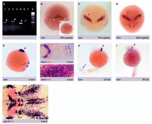
him is expressed dynamically during embryonic development, in exact overlap with her5 within the midbrain-hindbrain domain. (A) Maternal expression of him and her5, revealed by RT-PCR. (1,8). (1) him specific primers with cDNA isolated from four-cell stage embryos; (2) him specific primers without cDNA; (3) her5 specific primers with cDNA isolated from four-cell stage embryos; (4) her5 specific primers without cDNA; (5) pax2.1 specific primers with cDNA isolated from three-somite stage embryos; (6) pax2.1 specific primers with cDNA isolated from four-cell stage embryos; (7) pax2.1 specific primers without cDNA; (8) 100 bp DNA ladder (Fermentas MBI). Note the selective amplification of him and her5 in lanes (1) and (3) (white arrowheads), compared to the negative control pax2.1 (red arrowhead). (B-J) him expression revealed by whole-mount ISH (probe combination color-coded and indicated at the bottom left of each panel; stages at the bottom right; (B-D) dorsal views, anterior up; (E,H,I) lateral views, anterior left; (F,G,J) dorsal views of flat-mounted embryos, anterior left). At 30% epiboly (B), him is transcribed in the deep layer of the mesoderm (red arrows, see sagittal view in B′) and in scattered cells of the dorsal embryonic margin (white arrowheads). him expression within the MH domain (red arrowheads) is initiated at 75% epiboly (C) (note the difference in expression in medial and lateral parts of the IZ is indicated with white arrows) and maintained until 36 hpf (I). Note in F (and see higher magnification of the boxed area in G) that him and her5 expression in this domain are exactly coincident. him expression in the presomitic mesoderm starts at 90% epiboly (D) (blue arrows) and is maintained until 24 hpf (H). him and ngn1 are complementarily expressed in the MH region (J). Red arrowheads indicate him expression at the MHB and blue arrows expression in the presomitic mesoderm. IZ, intervening zone; vcc, ventrocaudal cluster, r2M, presumptive motorneurons of rhombomere 2; r2L, presumptive lateral neurons of rhombomere 2; r4M, presumptive motorneurons of rhombomere 4; r4L, presumptive lateral of rhombomere 4. |
|
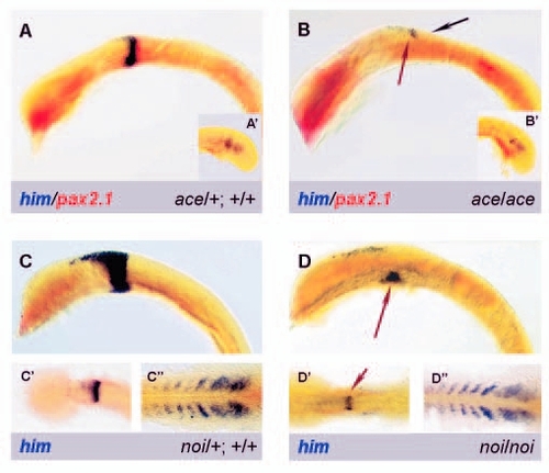
him expression is controlled by Pax2.1 and Fgf8 during the MH maintenance loop. (A,B) him (blue) and pax2.1 (red) expression in ace mutants (B) and WT siblings (A) at the 21-somite stage (all embryos deyolked, lateral view, anterior left). Note that both genes are coincidentally switched off at the MHB, except for a common dorsal patch (arrows), while him expression in the presomitic mesoderm is intact (A',B', insets). (C,D) him expression (blue) in noi mutants (D) and WT siblings (C) at the 17-somite stage (C,D: deyolked embryos, lateral views, anterior left; C'-D'': dorsal views of flat-mounted heads (C',D') and tails (C'',D''). him expression at the MHB is restricted to a ventral patch in noi (arrow), while presomitic expression is unaffected (C'',D''). EXPRESSION / LABELING:
|
|
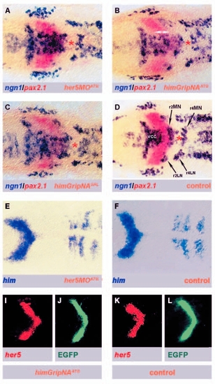
The activity of both Him and Her5 is necessary to prevent neurogenesis across the medial IZ in vivo. (A-D) The inhibition of either Him or Her5 function triggers ectopic neurogenesis in place of the MIZ (dorsal views of the MH region in flat-mounted embryos at the four-somite stage, anterior to the left). Embryos are probed for ngn1 (blue) and pax2.1 (red) expression following injection of her5MOATG(A), himGripNAATG (B), himGripNASPL (C) (orange labels), compared to a non-injected WT control embryo (D). Note that the vcc and r2MN are bridged by ectopic ngn1-positive cells (double arrows) after blocking Her5 or Him activity, while other undifferentiated areas are not affected (e.g. area between r2MN and r4MN, asterisk. (G-L) him and her5 expression are not successive and interdependent steps of the anti-neurogenic cascade acting in the MIZ (dorsal views of the MH area in flat-mounted embryos at the three-somite stage, anterior to the left, used markers are color-coded). (G,H) him expression in wild-type embryos (H) or after injection with her5MOATG (G). Note that him expression is not modified. (I-L) Expression of her5 (I,K) and GFP (J,L) in her5PAC::egfp embryos injected (K,L) or not (I,J) with himGripNAATG. Note that her5 and GFP expression are unaffected. vcc: ventro-caudal cluster, r2MN: prospective motorneurons of rhombomere 2, r4MN: prospective motorneurons of rhombomere 4. EXPRESSION / LABELING:
|
|
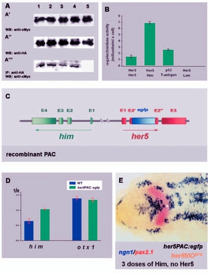
The crucial determinant of medial IZ formation is the level of Him + Her5 inhibitory activity, probably achieved in vivo by Him/Her5 heterodimers but replaceable by a higher level of either factor alone. (A) Co-immunoprecipitation assays reveal possible interactions between the bHLH transcription factors important to prevent/promote neurogenesis at the MHB. Crude protein extracts were isolated from yeasts transformed with the following constructs combinations: (1) her5pGBKT/ + her5pGADT7, (2) her5pGBKT7 + ngn1pGADT7, (3) himpGBKT7 + her5pGADT7, (4) T-antigen pGBKT7 + p53pGADT7; (5) her5pGADT7 + lampGADT7. Isolated extracts were either probed with anti-cMyc antibodies (A') and anti-HA antibodies (A'') or immunoprecipitated with anti-HA antibodies and then probed with anti-cMyc antibodies (A'''). (B) Stringency of Her5 homodimerization and Her5/Him heterodimerization, based on beta-galactosidase activity of yeast cells expressing appropriate construct combinations (Lazo et al., 1978). Note that the interaction between Him and Her5 is significantly stronger than Her5 homodimerization. (C-E) A higher dose of Him alone can compensate for the loss of Her5 activity and maintain the MIZ. (C) Schematic representation of the transgene integrated to generate her5PAC::egfp embryos (Tallafuss and Bally-Cuif, 2003): the egfp cDNA (blue cylinder) is inserted into the her5 region coding for the bHLH domain, resulting in a dysfunctional protein unable to bind both DNA and other bHLH factors. However, the him gene, contained in the PAC, is intact. (D) Quantification of him and otx1 (control) mRNAs in her5PAC::egfp transgenic compared to wild-type embryos using real-time RT-PCR. We do not know the number of recombined PAC copies integrated into the genome in our transgenic lines; however, note that the amount of him mRNA is 1.5-fold higher in the her5PAC::egfp transgenic embryo than in wild-type siblings. The change in threshold-crossing cycle (1/R) is shown for each mRNA relative to that for pax6 (assumed as a housekeeping gene) (a decrease in threshold-crossing corresponds to increase in mRNA level). The increase in him expression in the transgenic line is significant (P<0.02 by Student's t- test). Standard deviations are indicated with red lines. (E) Blocking Her5 activity (by injecting her5MOATG) in her5PAC::egfp transgenic embryos fails to trigger ectopic expression of ngn1 across the MIZ (white asterisk) (flat-mounted embryo at three somites, anterior left, used markers color-coded). |
|
The activity of both Her5 and Him is necessary to prevent neurogenesis in the lateral IZ. Dorsal views of flat-mounted -8.4ngn1::egfp transgenic embryos (Blader et al., 2003) probed at the three-somite stage for egfp expression (blue) after injection of 1 mM her5MOATG + 0.25 mM himGripNAATG (A), 1 mM her5MOATG (B), 0.25 mM himGripNAATG (C), compared to an uninjected control (D). A' and D' are enlargements of the boxed areas in A and D, respectively. The IZ is indicated by pax2.1 expression (red staining). Note that the LIZ is undergoing ectopic neurogenesis only in the embryo injected with both her5MOATG and himGripNAATG (blue arrows). EXPRESSION / LABELING:
|
|
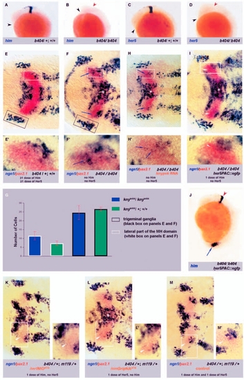
Ectopic neurogenesis in both the medial and lateral IZ in b404 deletion mutants results from the deletion of him and her5. (A-D) b404 mutants lack her5 and him expression. Lateral views of whole-mount embryos assayed for him or her5 expression at the 17-somite stage (anterior left, probes indicated bottom left, genotype bottom right). The position of the MHB in mutant embryos is indicated with a red arrowhead. The position of the head, reflecting the delayed convergence and extension problems in the mutant embryo, is indicated with a black arrowhead. (E-G) Ectopic neurogenesis across the LIZ revealed by ngn1 expression (blue) in b404 mutants (F,F') compared to non-mutant siblings (E,E'). In b404 embryos, ectopic ngn1-positive cells are present both in the lateral (blue arrow) and medial (asterisk) IZ (E' and F' are high magnification of the areas boxed in white in E and F, respectively). (G) The number of ngn1-positive cells in the future alar MH (area indicated with white box in E and F) is 40% higher in mutant embryos compared to WT siblings, while other neural plate areas are not affected (e.g. trigeminal ganglia, area boxed in black in E and F). (H) Reintroducing Knypek function in b404 mutants does not alter the IZ neurogenesis phenotype. Expression of ngn1 (blue) in three-somite b404 mutants where Kny function has been restored by kny RNA injection (dorsal views of flat-mounted embryos, anterior left, A' is a high magnification of the area boxed in A). Note that ectopic ngn1 expression both across the MIZ (asterisk) and LIZ (blue arrows) is not altered compared to uninjected b404 mutants (Fig. 7F). (I-J) Restoring Him function at endogenous levels in b404 mutants is sufficient to rescue the LIZ. (J) Crossing the b404 mutation into the her5PAC::egfp background generates b404/b404;her5PAC::egfp embryos where him expression is recovered with endogenous levels and expression pattern (lateral view of a 17-somite embryo, anterior left). (I,I') Expression of ngn1 (blue) in three-somite b404/b404;her5PAC::egfp embryos (dorsal views of flat-mounted embryos, anterior left, I' is a high magnification of the area boxed in I). Note that no ectopic neurogenesis is detectable any longer across the LIZ (white arrowheads), while the MIZ remains bridged by ectopic ngn1-positive cells (asterisk). (K-M). Him and Her5 equally contribute to the total inhibitory activity and one copy of either Him or Her5 is sufficient for formation of the LIZ. Formation of the LIZ in the b404/+; m119/+ embryos is indistinguishable in Him morphants (K,K'), Her5 morphants (L,L') and uninjected embryos (M,M') (white arrowheads). Note that after blocking either Him or Her5 activity ectopic neurogenesis occurs in the MIZ (white asterisk). K',L' and M' are enlargements of boxed area in K, L and M respectively (25 embryos was analyzed for b404/+; m119/+ injected with her5MOATG, 27 for b404/+; m119/+ injected with himGripATG and 20 uninjected b404/+; m119/+). EXPRESSION / LABELING:
|

