- Title
-
Evolution and expression of D2 and D3 dopamine receptor genes in zebrafish
- Authors
- Boehmler, W., Obrecht-Pflumio, S., Canfield, V., Thisse, C., Thisse, B., and Levenson, R.
- Source
- Full text @ Dev. Dyn.
|
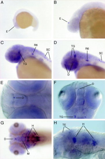
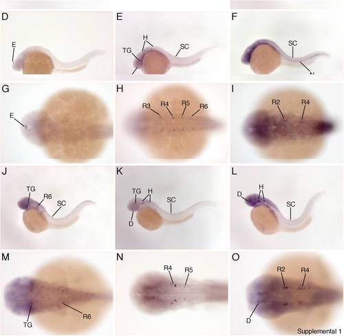
Expression of the dopamine receptor drd2a gene. A?D: Lateral views of embryos at mid-somitogenesis (15-somite stage, A), 24 hours postfertilization (hpf, B), 36 hpf (C), and 48 hpf (D). E,F: Differential interference contrast microscopy (DIC) images at 48 hpf. G: At 5 dpf (dorsal view). H: DIC image of a 5 dpf embryo. D, diencephalon; E, epiphysis; H, hindbrain; M, midbrain; R, rhombomere; SC, spinal cord; TE, tectum; TG, tegmentum. EXPRESSION / LABELING:
|
|
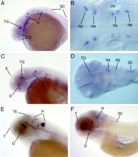
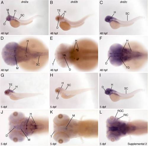
Expression of the drd2b gene. A: A 24 hours postfertilization (hpf) embryo (lateral view). B: Differential interference contrast microscopy (DIC) image of a 24 hpf embryo. C: At 36 hpf (lateral view). D: DIC image of a 36 hpf embryo. E: At 48 hpf (lateral view). F: 5 dpf (lateral view). D, diencephalon; E, epiphysis; H, hindbrain; R, rhombomere; SC, spinal cord; T, telencephalon; TE, tectum; TG, tegmentum. The unstained otic vesicle (OV) is shown for orientation. |
|
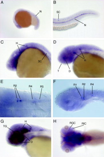
Expression of the drd2c gene. A: At mid-somitogenesis (15-somite stage, lateral view). B: At 24 hours postfertilization (hpf; tail, lateral view). C: At 24 hpf (head, lateral view). D: At 36 hpf (lateral view). E,F: Differential interference contrast microscopy (DIC) images of a 36 hpf embryo. G: At 48 hpf (lateral view). H: At 5 dpf (dorsal view). D, diencephalon; H, hindbrain; N, notochord; R, rhombomere; RGC, retinal ganglion cell layer; RIC, retinal intermediate cell layer; SC, spinal cord; T, telencephalon; TG, tegmentum. EXPRESSION / LABELING:
|
|
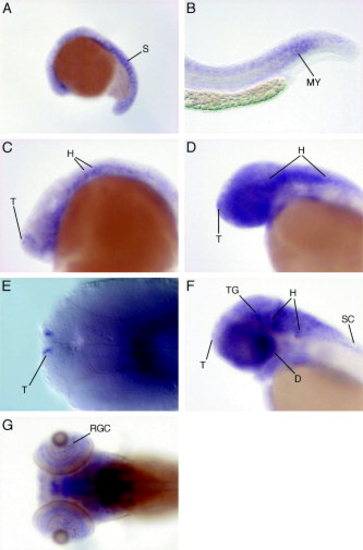
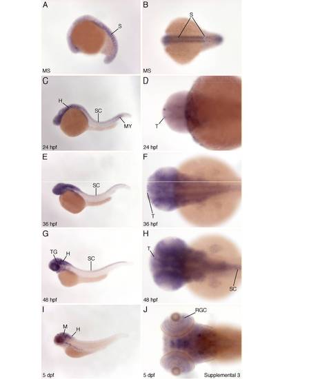
Expression of the dopamine receptor D3 gene drd3. A?D: Lateral views of embryos at mid-somitogenesis (A), 24 hours postfertilization (hpf; tail, B), 24 hpf (head, C), and 36 hpf (D). E: Differential interference contrast microscopy (DIC) image at 36 hpf. F: At 48 hpf (lateral view). G: At 5 dpf (dorsal view). D, diencephalon; H, hindbrain; MY, myotomes; RGC, retinal ganglion cell layer; S, somites; SC, spinal cord; T, telencephalon; TG, tegmentum. EXPRESSION / LABELING:
|
|
|
|
|
|
|

