- Title
-
A nonsense mutation in zebrafish gata1 causes the bloodless phenotype in vlad tepes
- Authors
- Lyons, S.E., Lawson, N.D., Lei, L., Bennett, P.E., Weinstein, B.M., and Liu, P.P.
- Source
- Full text @ Proc. Natl. Acad. Sci. USA
|
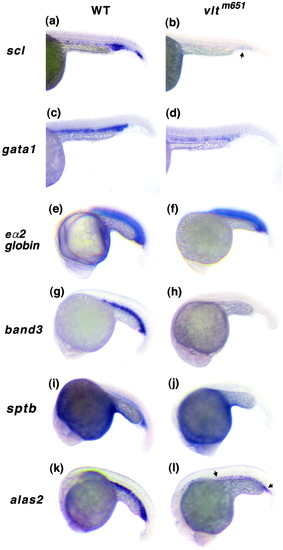
Expression of hematopoietic genes in vltm651 by whole-mount RNA in situ hybridization. Embryos from vltm651 heterozygote incrosses are shown in lateral views with the heads to the left. Embryo stages are 26 hpf (a-d) and 21 somites (e-l). In situ analyses were performed on embryos from at least three independent crosses. Wild-type sibling embryos (WT) are shown in a, c, e, g, i, and k, and mutant embryos (vltm651) are shown in b, d, f, h, j, and l with scl (a and b), gata1 (c and d), globin eα2 (e and f), band3 (g and h), sptb (i and j), and alas2 (k and l) RNA probes. An arrow in b shows residual staining in the posterior ICM with scl. Arrows in l demarcate the reduced staining in the ICM with alas2. |
|
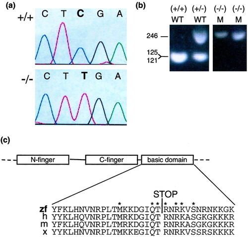
Detection of a nonsense point mutation in gata1. (a) The chromatograms show sequence of PCR products derived from a homozygous wild-type embryo (+/+) (Upper) and a homozygous mutant embryo (-/-) (Lower) for the gata1 gene nt 1013-1017. (b) Genotyping of vltm651 embryos. PCR products using primers Arg-339-S and Arg-339-AS synthesized from embryo DNA of vltm651 incrosses were digested with TaqI and electrophoresed on a 2% agarose gel. After TaqI digestion, mutant alleles appear as 246-bp and wild-type alleles as 121- and 125-bp products, which migrate together in the gel shown. Phenotypically wild-type embryos (WT) identified by presence of circulating blood and phenotypically mutant (M) embryos identified by absence of circulating blood are shown. (c) A schematic representation of the N-finger, C-finger, and basic domain of Gata1 and a sequence alignment of the basic domain from different species zebrafish (zf), human (h), mouse (m), and Xenopus (x) are shown with the Arg-339 → Stop mutation identified by a STOP. The asterisks mark the aminoacids that make direct contact with the minor groove of DNA (29). |
|
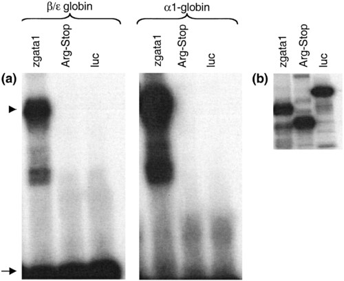
DNA-binding activity of Gata1 and Gata1 (Arg-339 → STOP). (a) Unlabeled in vitro translated proteins, wild-type zebrafish Gata1 (zgata1), zebrafish Gata1 (Arg-339 → Stop) (Arg-Stop), and negative control protein, luciferase (luc), were used in an electrophoretic mobility shift assay with the indicated 32P-labeled probes, β/ε globin or α1-globin. The DNA-protein complexes are seen as slower migrating bands, marked by an arrowhead. The arrow marks free probe, demonstrating excess of DNA in the mixture. (b) Polyacrylamide gel electrophoresis of in vitro translated proteins generated in the presence of [35S]methionine and visualized by autoradiography. |
|
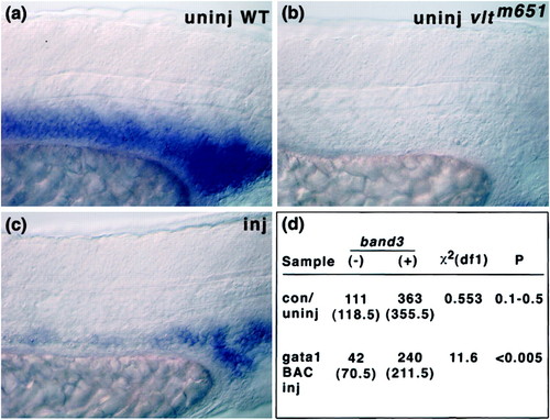
Phenotypic rescue of vltm651 embryos by injection of gata1-BAC DNA. (a-c) Lateral views of the ICM regions of embryos stained by in situ hybridization with band3 (x400 magnification). (a) Uninjected phenotypically wild-type embryo (band3-positive), (b) uninjected phenotypically mutant embryo (band3-negative), (c) gata1-BAC injected embryo showing mosaic expression of band3. (d) χ2 analysis of injection data. The null hypothesis tested was that injections did not change the inheritance of the mutant phenotype (i.e., recessive or 1:3 mutant to wild-type ratio). The data shown indicate the number of embryos obtained for each phenotype, and the numbers shown in parentheses are the number of embryos expected for each phenotype assuming recessive inheritance. |


