- Title
-
Sonic hedgehog is required early in pancreatic islet development
- Authors
- diIorio, P.J., Moss, J.B., Sbrogna, J.L., Karlstrom, R.O., and Moss, L.G.
- Source
- Full text @ Dev. Biol.
|
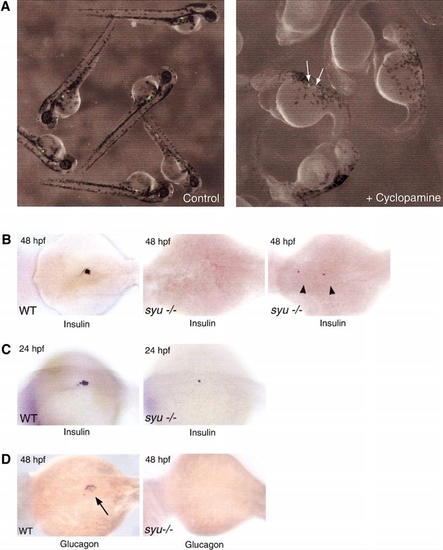
Disruption of Shh signaling alters pancreas development. (A) Homozygous 48-hpf INS-GFP transgenic zebrafish. Untreated embryos (left) contain a compact multicellular cluster of GFP-expressing cells. Addition of 50 μM cyclopamine at 4 hpf (right) blocks formation of GFP+ islets. Some isolated, single GFP-expressing cells remain (arrows). (B) 48 hpf WT zebrafish in situ hybridized as whole-mount embryos with a proinsulin riboprobe depict normal clusters of insulin-expressing cells in the pancreas budding from the right side of the endoderm (left). 48 hpf Syut4-/- embryos have lost all insulin expression (middle) or exhibit expression (right) in isolated single cells (arrowheads). (C) Early insulin expression in a 24-hpf WT (left) and syu embryo (right). (D) Hybridization of WT embryos with a proglucagon probe (left) reveals cells in the perimeter of the islet as well as in an isolated cell within the gut at 48 hpf (arrow). No mRNA for this neuroendocrine hormone is seen in syu embryos (right). (B?D) Views are dorsal, with anterior to the left. |
|
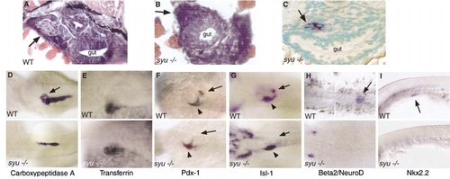
Nature of the islet defect in the zebrafish mutant syut4. The morphology of WT (A) vs syu embryos (B) was compared. Paraffin transverse cut sections (5 μm), stained with hematoxylin and eosin. (A) WT islet (arrow). (B) The islet is selectively missing in syu embryos (arrow). (C) Carboxypeptidase A expression (arrow) in 72-hpf syu embryos embedded in paraffin, sectioned, and counterstained with Vector Green. (D?I) Whole-mount in situ hybridization analysis of Wt and syu-/- embryos. Views are dorsal with anterior to the left, except (I), which is a lateral view. (D) Carboxypeptidase A expression in 48-hpf WT and syu-/- embryos. (E) Expression 72 hpf of transferrin (F) Pdx-1 expression in 48-hpf WT pancreas (arrow) and duodenum (arrowhead). (G) Isl-1 expression in 48-hpf syu-/- embryos. Arrows denote loss of expression. Foregut expression (arrowheads) is retained. (H) Loss of Beta2/NeuroD expression within the islet at 48 hpf (arrow) in syu-/- embryos. (I) Nkx2.2 (nk2.2) expression in WT 24-hpf embryos in early gut endoderm where the pancreatic diverticulum will form (arrow). |
|
Insulin expression in hh signal transduction mutants. Insulin expression in 48-hpf WT (A), homozygous yotty17a-/- (B) and smub577-/- embryos (C). Arrows denote the presence of insulin-labeled cells in the mutants (B, C). |
|
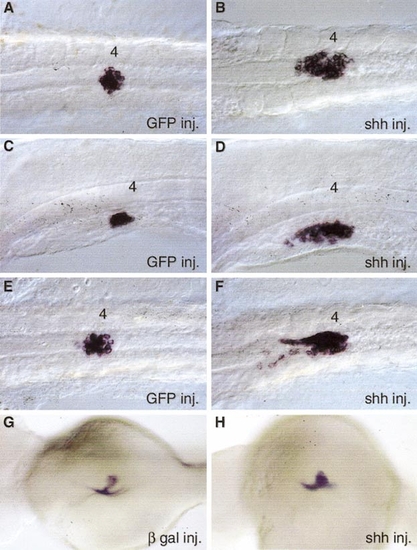
Insulin and Pdx-1 expression after shh overexpression in WT embryos. Left: mRNA control-injected embryos. Right; shh mRNA-injected embryos. After mRNA injection at the two- to four-cell stage, embryos developed for 24 or 48 hpf and were subjected to whole-mount in situ analysis using the insulin or Pdx-1 probe. (A, C, E) Control GFP mRNA-injected embryos hybridized at 24 hpf with the insulin probe. Positive cells are centered ventral to somite 4 (4). (B, D, F) Shh mRNA-injected embryos hybridized with the insulin probe. Note the anterior extension of labeled cells. (G) Control β-gal mRNA-injected embryo hybridized with the Pdx-1 probe. (H) Shh mRNA-injected embryo hybridized with the Pdx-1 probe. (A, B, E, F) Ventral views. (C, D) Lateral views. (G, H) Dorsal views. |
|
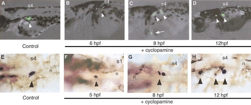
Shh signaling is required early for patterning the endocrine islet. Living fifth-generation homozygous INS-GFP or WT fixed embryos imaged at 48 hpf untreated (A, E) or treated with 50 μM cyclopamine at 6 (B, F), 8 (C, G), or 12 (D, H) hpf. Representative islet phenotypes are shown in living embryos as fluorescent (A?D) or as bright-field images after fixation and in situ hybridization with insulin probe (E?H). (A) 48-hpf untreated INS-GFP embryos. (B) Exposure to cyclopamine at 6 hpf. (C) Treatment at 8 hpf. (D) Cyclopamine exposure at 12 hpf. (E) Endogenous insulin expression in control ethanol-treated embryos. (F?H) Cyclopamine-treated embryos. Cyclopamine was added at 5 hpf (F); branchial arches (arrowhead), 8 (G) and 12 hpf (H). Abbreviations: s4, somite 4; s1, somite1; n, notochord; arrows, position of INS-GFP fluorescent cells; arrowheads, position of insulin-expressing cells. |
Reprinted from Developmental Biology, 244(1), diIorio, P.J., Moss, J.B., Sbrogna, J.L., Karlstrom, R.O., and Moss, L.G., Sonic hedgehog is required early in pancreatic islet development, 75-84, Copyright (2002) with permission from Elsevier. Full text @ Dev. Biol.


