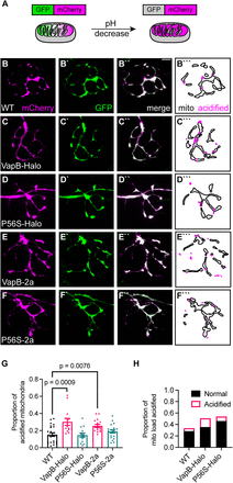
Fig. 3 VapB expression increases mitophagy in the axon terminal. A, Schematic of mitophagy indicator mito-GFP-mCherry localization and fluorescence in the mitochondrion. In low pH, such as in a lysosome, the GFP is quenched. B?D, Representative images of axon terminals expressing the mitophagy indicator in WT (B), VapB-HaloTag transgenic (VapB-Halo; C), VapBP56S-HaloTag transgenic (P56S-Halo; D), VapB-p2a-TagBFP transgenic (VapB-2a; E), and VapBP56S-p2a-TagBFP transgenic (P56S-2a; F). Distribution of acidified (magenta) and unacidified (outline) mitochondria is illustrated (B```?F```). G, Proportion of acidified mitochondria relative to the total mitochondrial population. H, Proportion of mitochondrial load acidified (mCherry-only mitochondrial area/average mitochondrial axon terminal load derived from Fig. 1H). Scale bar, 5 ?m in B``. Each data point for G represents the average calculated from an individual animal. All data represented as mean ± SEM. ANOVA with Dunnett's post hoc contrasts in G.
Image
Figure Caption
Acknowledgments
This image is the copyrighted work of the attributed author or publisher, and
ZFIN has permission only to display this image to its users.
Additional permissions should be obtained from the applicable author or publisher of the image.
Full text @ J. Neurosci.

