
|
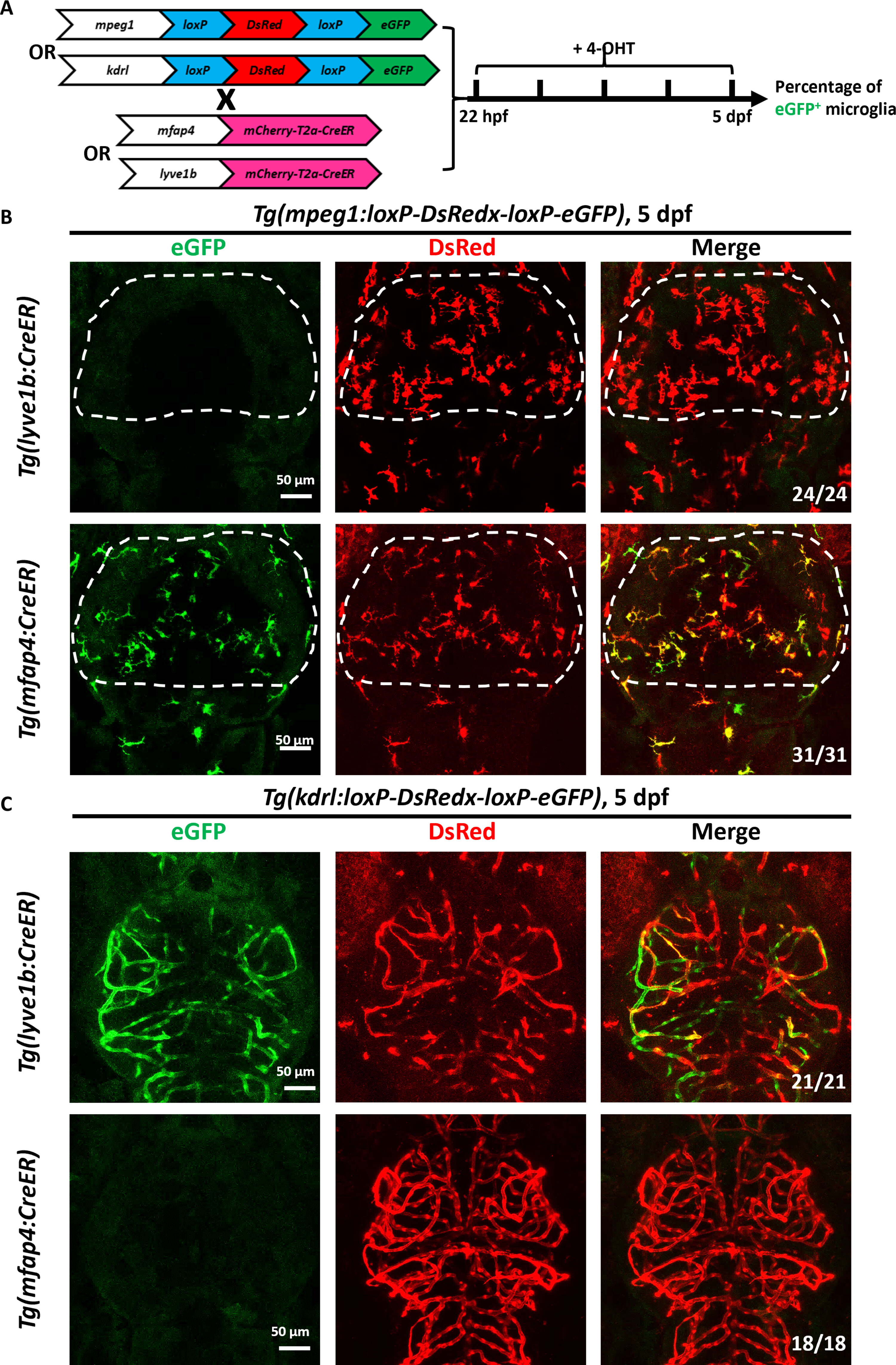
Fig. 3 Microglia originated from macrophages instead of lyve1b+ lymphatic vessels. (A) Schematic view of lineage tracing workflow. (B) Fluorescent images show the labeling efficiency of GFP+ microglia in the brain of 5-dpf Tg(lyve1b:CreER;mpeg1:loxP-DsRedx-loxP-eGFP) and Tg(mfap4:CreER;mpeg1:loxP-DsRedx-loxP-eGFP) larvae treated with 4-hydroxytamoxifen (4-OHT) from 22 hpf to 5 dpf. (C) Fluorescent images show the labeling efficiency of GFP+ blood vessels in the brain of 5-dpf Tg(lyve1b:CreER;kdrl:loxP-DsRedx-loxP-eGFP) and Tg(mfap4:CreER;kdrl:loxP-DsRedx-loxP-eGFP) larvae treated with 4-OHT from 22 hpf to 5 dpf.