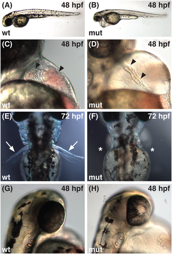
Fig. 1 The sk29 mutation causes multiple embryonic defects. (A, B) Lateral views of live embryos at 48 hpf, anterior to the left, dorsal to the top. In comparison to wild-type embryos (wt), sk29 mutant embryos (mut) have a slightly shorter body axis, decreased pigmentation, pericardial edema, and a thickened yolk extension. (C, D) Lateral views at 48 hpf, anterior to the top, dorsal to the left. In sk29 mutants, cardiac chambers (arrowheads) fail to expand and remain relatively linear, with little apparent morphological distinction of the AVC. (E, F) Dorsal views at 72 hpf, anterior to the top. Pectoral fins (arrows) are absent (asterisks) in sk29 mutants. (G, H) Lateral views at 48 hpf, anterior to the top, dorsal to the left. Retinal pigmentation is abnormal in sk29 mutants.
Image
Figure Caption
Figure Data
Acknowledgments
This image is the copyrighted work of the attributed author or publisher, and
ZFIN has permission only to display this image to its users.
Additional permissions should be obtained from the applicable author or publisher of the image.
Full text @ Dev. Dyn.

