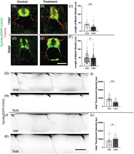
Fig. 4 Regeneration of myelinated segments following recovery from acute hyperglycemia.(A & B) Representative images of C12 and Tx12 Tg(mbp:EGFP-CAAX) zebrafish labeled with antibodies against GFP (green) and acetylated ?-tubulin (red) following the treatment schedule described in Figure 1A. (C) The length of myelinated segments were significantly shorter in Tx12 fish compared to C12 fish, ****P < .0001. (D and E) Representative images of C22 and Tx22 Tg(mbp:EGFP-CAAX) zebrafish labeled with antibodies against GFP (green) and acetylated ?-tubulin (red) following the treatment and recovery schedule described in Figure 2A. (F) Following the 10-day recovery period, no difference was observed in the length of myelinated segments between Tx22 and C22 fish, P = .4413. (G and H) Lateral view of C12 and Tx12 myelinated segments. (I) 7-day incubation in 120 mM D-glucose solution significantly diminished mbp+ fluorescent intensity in Tx12 fish compared to C12, ****P < .0001. (J and K) Lateral view of C22 and Tx22 myelinated segments. (L) Following recovery, Tx22 myelinated segments had a significantly greater mbp+ fluorescent intensity compared to C22, **P = .0055. au, arbitrary units. All values are means ± SEM. Scale bars = 50 ?m.
Image
Figure Caption
Acknowledgments
This image is the copyrighted work of the attributed author or publisher, and
ZFIN has permission only to display this image to its users.
Additional permissions should be obtained from the applicable author or publisher of the image.
Full text @ Dev. Dyn.

