
Fig. 1
|
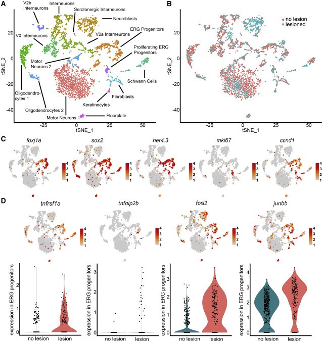
Fig. 1 scRNA-seq indicates importance of TNF signaling in progenitor cells (A) Unsupervised clustering of her4.3:GFP+ cells identifies progenitor and other neural clusters. (B) Cells from lesioned and unlesioned samples contribute to cell clusters with a disproportionally high contribution of the lesioned sample to ?neuroblasts.? (C) Marker gene distribution identifies proliferating ERG progenitor cell clusters. (D) TNF receptor (tnfrsf1a), downstream gene (tnfaip2b), and components of AP-1 signal transducing complex (fosl2, junbb) are enriched in ERG progenitor cells (tSNE plots), and expression is increased after injury (violin plots). For clarity, only cells with expression of > 2 are indicated in the feature plot of junbb. Color codes indicate expression levels from low (gray) to high (red) (see also Figure S1; Table S1).
Reprinted from Developmental Cell, 56(11), Cavone, L., McCann, T., Drake, L.K., Aguzzi, E.A., Opri?oreanu, A.M., Pedersen, E., Sandi, S., Selvarajah, J., Tsarouchas, T.M., Wehner, D., Keatinge, M., Mysiak, K.S., Henderson, B.E.P., Dobie, R., Henderson, N.C., Becker, T., Becker, C.G., A unique macrophage subpopulation signals directly to progenitor cells to promote regenerative neurogenesis in the zebrafish spinal cord, 1617-1630.e6, Copyright (2021) with permission from Elsevier. Full text @ Dev. Cell