IMAGE

Fig. 2

ID
ZDB-IMAGE-210403-69
Genes
Source
Figures for Kesavan et al., 2020
Image
Figure Caption

Fig. 2

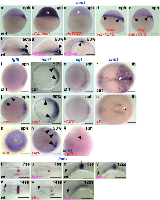
Ism1 expression depends on dorsal determinants, and Nodal and Fgf signaling. Misexpression of constitutively active β-catenin induces ectopic ism1 expression b, asterisk compared with controls a; c conversely, dominant negative TCF3 diminishes ism1 expression; However, bozd and sqte can induce ism1 (arrows) in the absence of Wnt/β -catenin signaling. f, gism1 expression in the blastoderm (arrows) is absent in MZoep mutants, while its expression in the YSL remains unaffected (asterisks). h Residual ism1 expression in MZoep mutants at the end of gastrulation (arrowhead). Both bozj and sqtk elicit ectopic fgf8 expression (arrows, asterisk) compared with controls i. Both sqtm and cycn elicit ectopic ism1 expression compared with controls l. oq Neither sqtp nor ism1 are ectopically induced by fgf8 overexpression. rz Fgf-dependence of ism1; r, s Pharmacological FgfR inhibition during gastrulation abolishes ism1 expression in the axial mesoderm and in the posterior paraxial mesoderm (white asterisks); v, w FgfR inhibition during somitogenesis abolishes ism1 expression in the MHB (red asterisks) and forebrain (arrowheads); (t,u,x,z) ism1 expression in the MHB (asterisk) and forebrain (arrowhead) is missing in fgf8/ace mutants. Note that expression in the MHB in panels xz appears to depend on the genetic dose of fgf8. Genotypes (pink), stages (black), treatment (red), and riboprobes (blue) are indicated on the panels. a-g Lateral views, dorsal to the right; h, xz lateral views, anterior to the left; ip animal views, dorsal to the right if discernible; q dorsal view, animal pole to the top; rw dorsal views, anterior to the left. Scale bar: 200 μm in ae and iq; 100 μm in fh and rz

Figure Data
Acknowledgments
This image is the copyrighted work of the attributed author or publisher, and ZFIN has permission only to display this image to its users. Additional permissions should be obtained from the applicable author or publisher of the image. Full text @ Cell Tissue Res.