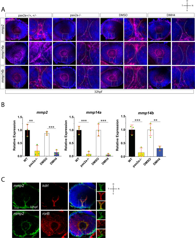
Hyaloid vasculature is a source of mmp2 during optic fissure fusion. (A) Fluorescent whole mount in situ hybridization comparing mmp2, mmp14a and mmp14b expression in WT vs pax2a−/− and DMSO vs DMH4 treated embryos at 32 hpf. Broken white lines outline the OF retinal lobes. mmp2, 14a and 14b expression within the OF is reduced in pax2a−/− and DMH4 treated embryos. Scale bar = 50 μm. (B) qPCR analysis confirms a decrease in expression of mmp2, 14a and 14b in both pax2a−/− and DMH4 treated embryos. (C) Two color whole mount in situ hybridization simultaneously examining mmp2 and kdrl, or mmp2 and rorB expression at 32 hpf. DNA was stained with DAPI. Scale bar = 50 μm. Clear overlap of signal is observed for mmp2 and kdrl, but not mmp2 and rorB.
This image is the copyrighted work of the attributed author or publisher, and
ZFIN has permission only to display this image to its users.
Additional permissions should be obtained from the applicable author or publisher of the image.
Full text @ Sci. Rep.
Your Input Welcome
Thank you for submitting comments. Your input has been emailed to ZFIN curators who may contact you if
additional information is required.
Oops. Something went wrong. Please try again later.