Fig. 4
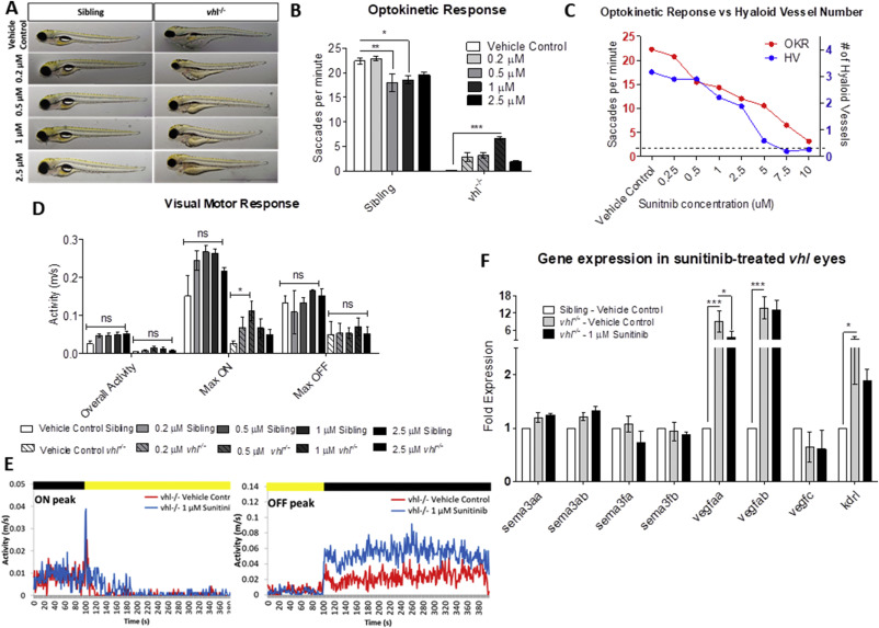
Fig. 4 Sunitinib malate significantly improves visual function and reduces VEGF expression in vhl?/? larvae. A) Gross morphology of sibling and vhl-/- larvae treated with increasing doses of sunitinib malate at 5 dpf. Treatment reduces yolk and cardiac oedema but does not result in swim bladder inflation. B) All concentrations of sunitinib tested increase OKR visual function at 5 dpf with 1??M sunitinib malate highlighting a statistically significant improvement. n?=?3, 12 technical replicates per experiment. C) Correlation between hyaloid vessel number and visual function in 5 dpf wildtype larvae treated with sunitinib malate. n?=?3 with 10 technical replicates per experiment. Dotted line denotes vhl?/? visual function and number of primary hyaloid branches at 5 dpf. D) VMR Max ?ON? response is significantly increased dose dependently in larvae treated with 0.2??M and 0.5??M Sunitinib. n?=?3, 12 technical replicates per treatment group per experiment. E) VMR chromatograms illustrating the average Max ON and Max OFF peaks after treatment. F) Expression of pro- and anti-angiogenic vhl?/? larval eyes following sunitinib treatment.
Reprinted from Developmental Biology, 457(2), Ward, R., Ali, Z., Slater, K., Reynolds, A.L., Jensen, L.D., Kennedy, B.N., Pharmacological restoration of visual function in a zebrafish model of von-Hippel Lindau disease, 226-234, Copyright (2019) with permission from Elsevier. Full text @ Dev. Biol.

