
|
Fig. 5
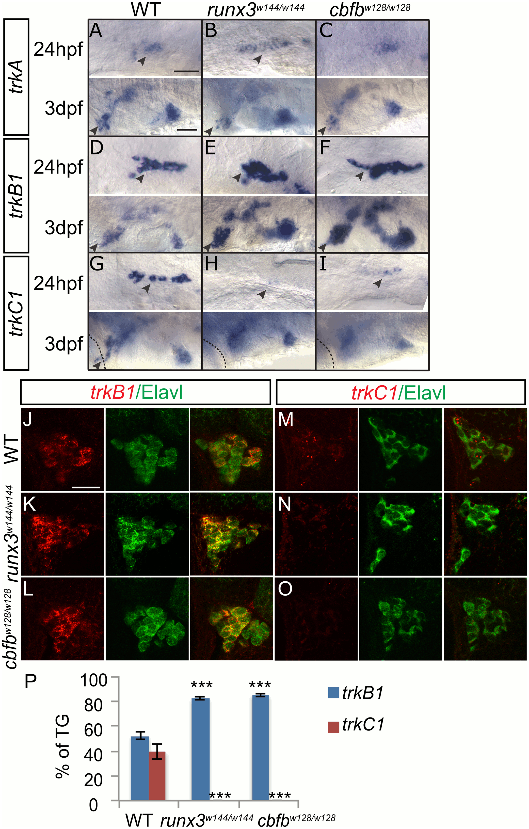
Loss of runx or cbfb expression affects trk receptor expression in the TG.
A-I, Colormetric in situ hybridization for trkA (A-C), trkB1 (D-F), and trkC1 (G-I) in the runx3w144/w144 and cbfbw128/w128 mutants focusing on the TG at 24hpf and 3dpf. J-O, Antibody staining of Elavl (green) in conjunction with fluorescent in situ hybridization for trkB1 (J-L) and trkC1 (M-O) in the runx3w144/w144 and cbfbw128/w128 mutants at 3dpf. P, Quantification of marker gene expression as total number of TG neurons/ganglion and as % TG at 3dpf. Arrowhead, TG; X, vagal ganglion. Scale bar: A-I 100 ?m, J-O 20?m. Embryos per condition (n = 3?5). ***p<0.001. All error bars represent S.E.M.