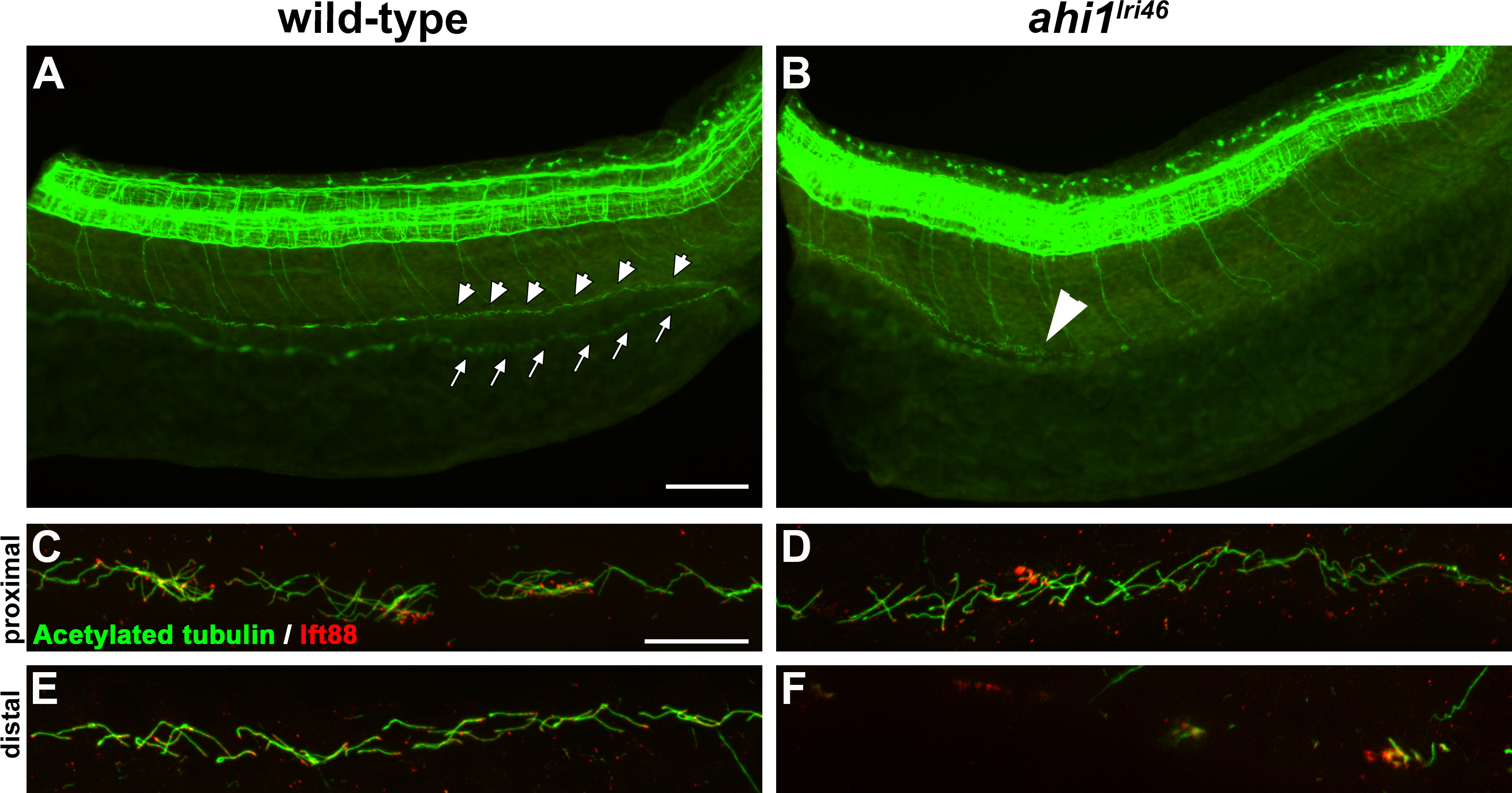
Fig. 2
Loss of ahi1 affects other ciliated tissues. (A, B) Anti-acetylated tubulin whole-mount immunostaining (green) of kidney duct in wild-type and ahi1lri46 mutants at 36 hpf. Anterior is to the left in both images. Cilia line the pronephric duct (small arrowheads) in wild-type animals. The contralateral duct also can be observed (small arrows). Cilia are absent in the middle and distal pronephros in ahi1lri46 mutants (arrowhead in [B] indicates the proximal boundary where cilia are missing). (C, D) In the proximal region, mutant cilia are not qualitatively different from wild-type controls. (E, F) Cilia in the distal region of the pronephros are absent in mutant animals when compared to controls. Ift88 immunostaining (red) was used to mark the ciliary transition zone. Scale bars: 50 μm (A, B) and 20 μm (C–F).

