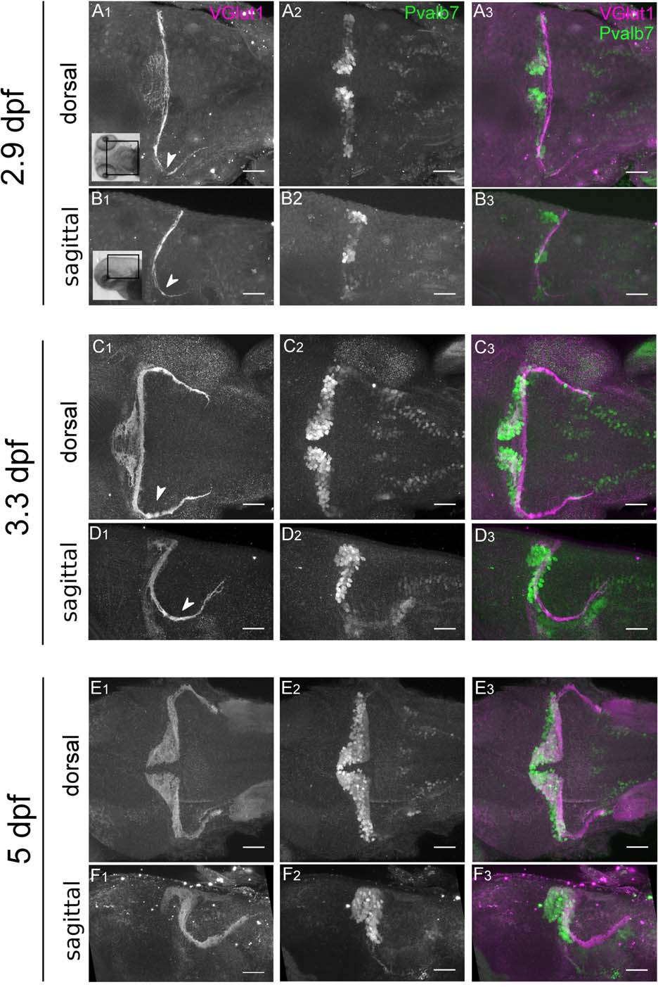
Fig. 6
Vglut1 expression in parallel fibers moves anteriorly early in development and coincides with Purkinje cell development. (A, B) Expression at 2.9 dpf (70 hpf) of VGlut1 (A1, B1) and Pvalb7 (A2, B2), in both dorsal and sagittal views. Merge (A3, B3) shows that VGlut1 and Pvalb7 show little overlap at this early time point. Additionally, VGlut1 expression can be found in parallel fibers that project posteriorly (A1, B1; white arrowheads). (C, D) Expression at 3.3 dpf (80 hpf) of VGlut1 (C1, D1), Pvalb7 (C2, D2) and merge (C3, D3) shows that parallel fibers expand forward, beginning to overlap with Purkinje cells during this time period. There is substantial outgrowth of parallel fibers projecting posteriorly (C1, D1; white arrowheads). (E, F) Expression at 5dpf shows that by this time point VGlut1 (E1, F1) and Pvalb7 (E2, F2) are highly spatially coordinated (merge; E3, F3) and both layers have begun to curve anteriorly. Scale bar 50 µm. Images taken using 20× (1.0 NA) water objective.

