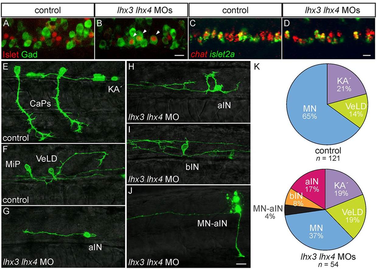
Fig. 5
Lhx3 and Lhx4 prevent PMNs from acquiring KA2 IN characteristics. (A-D) Lateral views of 24hpf embryos. (A,B) Islet and Gad are co-expressed in lhx3+lhx4 MO-injected embryos (arrowheads). (C,D) MNs continue to express chat in lhx3+lhx4 MO-injected embryos. (E-I) Mosaically labeled pMN derivatives in 28-32hpf s1020t embryos. (E,F) Injection of UAS:EGFPCAAX DNA labels PMNs, including both CaP (E) and MiP (F), as well as KA2 (E) and VeLD (F) in control embryos. (G-I) Mosaically labeled pMN derivatives in 28-32hpf s1020t embryos injected with lhx3 and lhx4 MOs. (G) Aberrant neuron with an axon that initially projects caudally before turning laterally and ascending the spinal cord (aIN; n=9). (H) Aberrant neuron with axon that ascends without a proximal descending segment. (I) Aberrant neuron with bifurcating axons, one ascending and one descending (bIN; n=4). (J) Dye-labeled MN-IN hybrid neuron with both ascending IN and peripheral MN axons in embryo injected with lhx3+lhx4 MOs (n=2); dye dorsal to the labeled neuron is from an adjacent cell killed during labeling. (K) Relative proportions of labeled pMN-derived neurons. Three new neuron classes appear in lhx3+lhx4 MO-injected embryos; only MNs are under-represented relative to control embryos (P=2.2×10⁵). The percentage of labeled cells is listed for each condition. Scale bars: 20µm.

