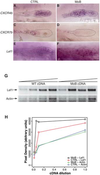
Fig. 5 Primordium patterning is altered in itchb knocked-down embryos.
(A?F) RNA in situ hybridization of factors required for primordium patterning in control embryos (vehicle-injected siblings) (CTRL, left panels) and MOs-injected embryos (MoB, right panels) at 30 hpf. cldnb:gfp embryos were used to ensure that the primordium had migrated pass the 10th somite before fixation. (A,B) cxcr4b expression was limited to the leading half of the primordium in control embryos, but extended throughout the primordium after itchb knockdown. (C,D) cxcr7b was limited to the trailing end of control embryos, and almost completely excluded from the primordium in itchb knockdown. (E,F) lef1 was expressed in the leading edge of control embryos. Darker staining indicated higher expression in embryos injected with MOs against itchb (MoB), but leading edge expression was maintained. Scale bar: 25 Ám. (G) Semi-quantitative RT-PCR showing enhanced expression of lef1 in embryos injected with MOs against itchb (MoB) as compared to control embryos (WT). Amplification of the actin gene was used as an internal control. The image was inverted to facilitate quantification. (H) Densitometry measurements from the gel presented in G. This is representative of three different experiments.

