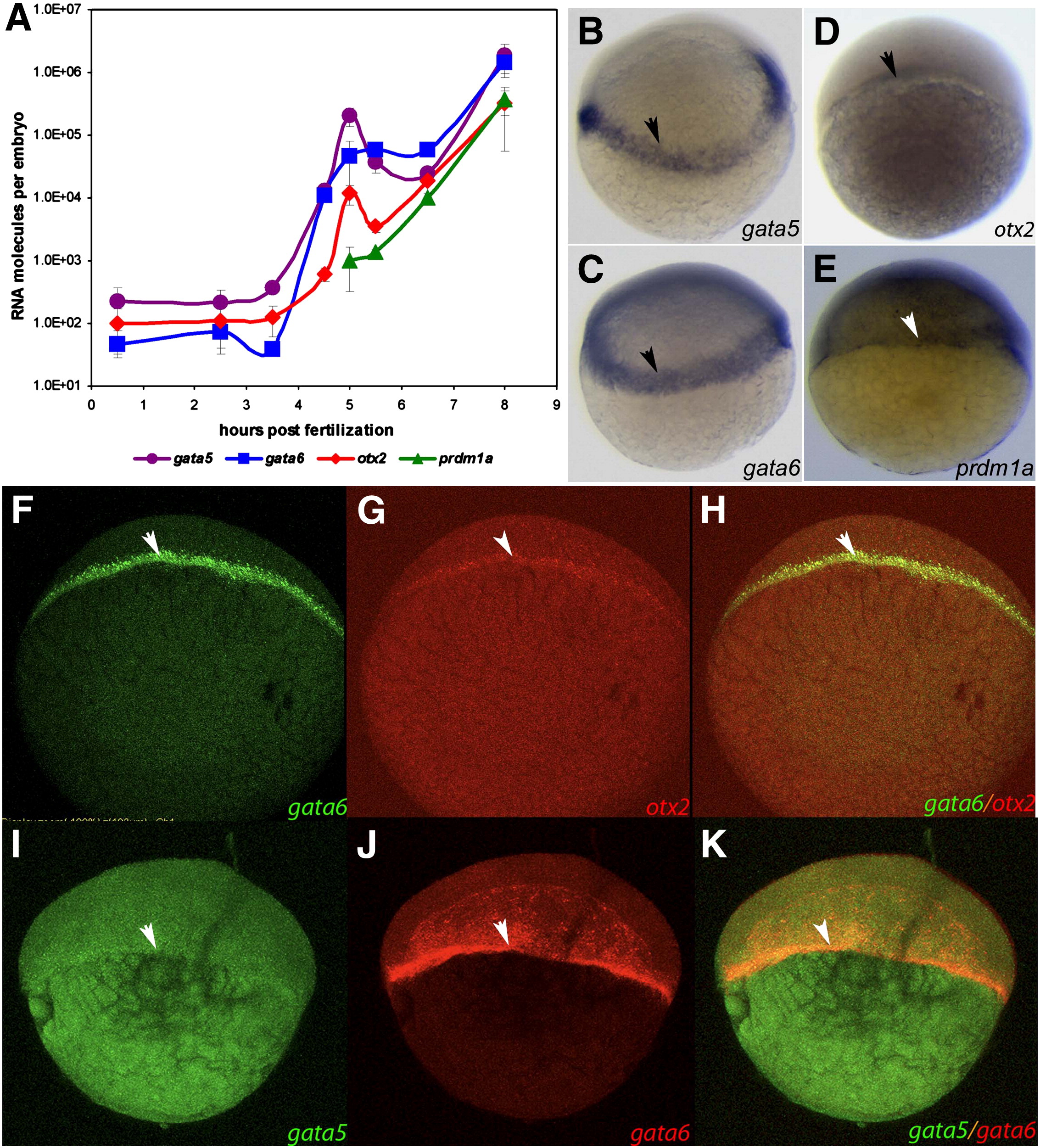
Fig. 1 Temporal and spatial expression profiles of gata5, gata6, otx2 and prdm1a. (A) Quantitative expression profiles of gata5, gata6, otx2 and prdm1a determined by using Q-PCR. The X-axis represents RNA molecules per embryo defined by Q-PCR. RNA was isolated from embryos at different time points, and the absolute molecular numbers per embryo are shown as a purple line for gata5, a blue line for gata6, a red line for otx2, and a green line for prdm1a. The experiments were conducted in triplicate, and the standard deviation is shown as a bar extending to both sides of the mean. (B?E) In situ hybridization of gata5, gata6, otx2 and prdm1a in wild-type embryos at 5 hpf. gata5 and gata6 are expressed in the mesendoderm and the YSL at 5 hpf. otx2 is expressed in the blastoderm at 5 hpf, and prdm1a is expressed in the blastoderm and ectoderm. Arrow indicates the location of mesendoderm lineage at the margin of blastoderm. (F?K) Co-localization of otx2, gata5 and gata6 at 5 hpf double in situ hybridization for gata6 and otx2 (F, G, H) and gata5 and gata6 (I, J, K) in wild-type embryos at 5 hpf. (F) gata6 mRNA is indicated by green fluorescence. (G) otx2 mRNA is shown by red fluorescence. (H) Merged image of F and G, indicating the co-localization of gata6 and otx2 in the mesendoderm. (I) gata5 mRNA is indicated by green fluorescence. (J) gata6 mRNA is shown by red fluorescence. (K) Merged image of I and J indicating the co-localization of gata5 and gata6 in the mesendoderm. Arrow indicates the location of mesendoderm lineage at the margin of the blastoderm.
Reprinted from Developmental Biology, 357(2), Tseng, W.F., Jang, T.H., Huang, C.B., and Yuh, C.H., An evolutionarily conserved kernel of gata5, gata6, otx2 and prdm1a operates in the formation of endoderm in zebrafish, 541-57, Copyright (2011) with permission from Elsevier. Full text @ Dev. Biol.

