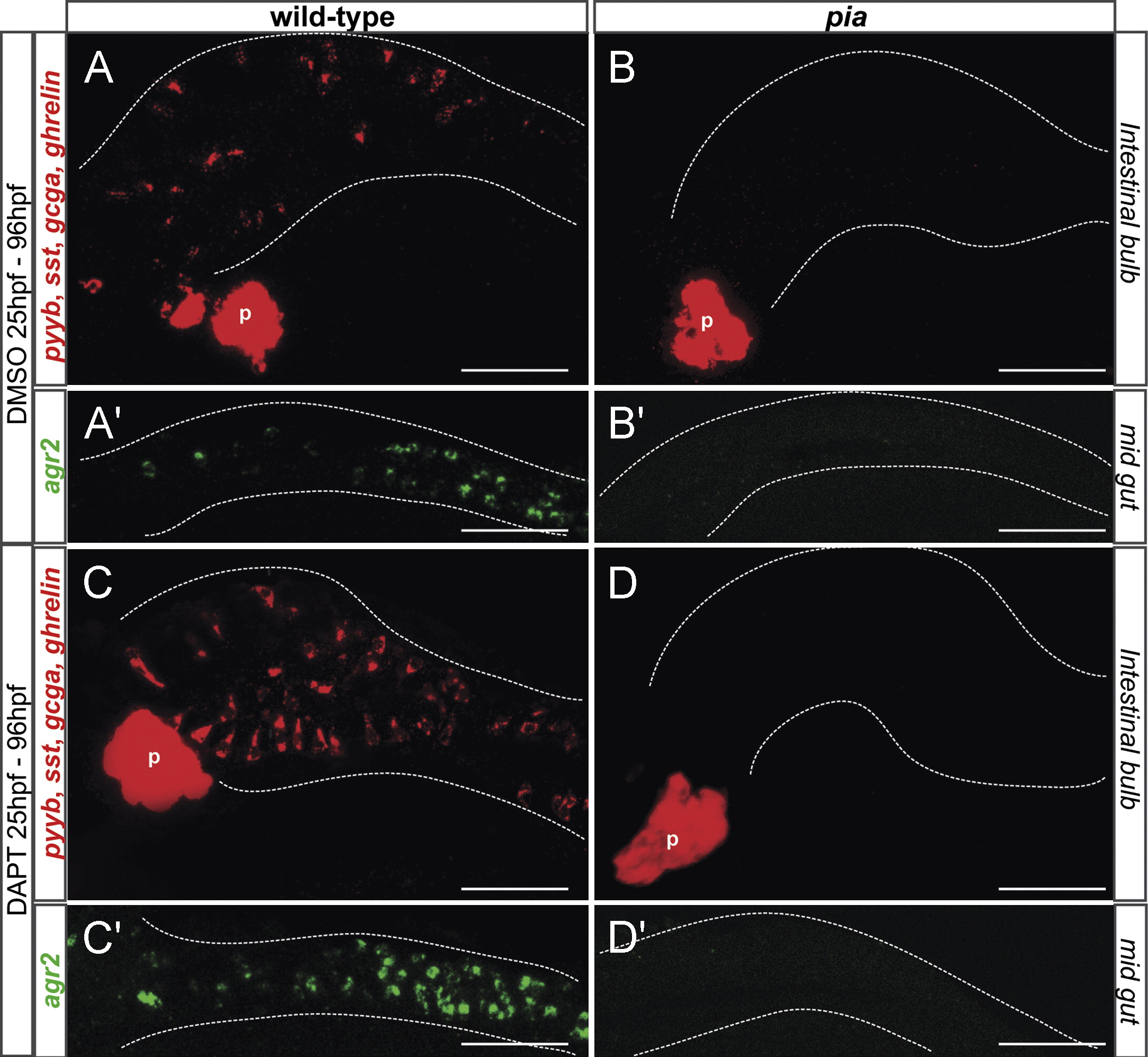
Fig. 6 Ascl1a is required for the induction of secretory cell fate upon inhibition of the Notch pathway. Ventral views with anterior to the left of embryos analyzed by double fluorescent WISH using a agr2 probe (green) and with a mixture of gcga, sst and ghrelin probes (red). All pictures are confocal projections. (A and C) WT embryos treated with DAPT from 25 to 96 hpf show an increased number of enteroendocrine and goblet cells (fold increase of 2.2±0.5 and 1.5±0.4, respectively). (B and D) pia embryos treated with DMSO or DAPT from 25 to 96 hpf are devoid or secretory cells in the gastrointestinal tract. P, pancreas. Scale bars: 50 μM.
Reprinted from Developmental Biology, 376(2), Flasse, L., Stern, D.G., Pirson, J.L., Manfroid, I., Peers, B., and Voz, M.L., The bHLH transcription factor Ascl1a is essential for the specification of the intestinal secretory cells and mediates Notch signaling in the zebrafish intestine, 187-197, Copyright (2013) with permission from Elsevier. Full text @ Dev. Biol.

