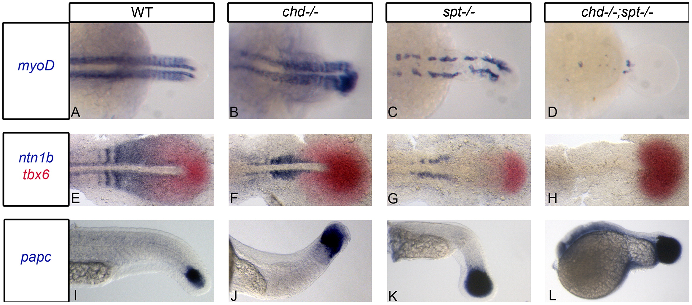
Fig. 2 Chd;spt mutants produce mesodermal progenitor cells in the tailbud but are not able to form somites. (A)–(D) Dorsal view of myoD expression in 24 hpf embryos. (A)–(C) WT, chd, and spt embryos form organized somites even if it is only in the tail (spt). chd;spt mutants do not form organized somites. (60/65 have no organized somites. The remaining 5 only had 2–3 somites form.) (D). (E)–(H), dorsal view of ntn1b expressing muscle cells and tbx6 expressing MPCs in 11 hpf embryos. (E)–(G) WT, chd, and spt embryos have MPCs in tailbud as well as differentiated muscle cells outside the tailbud. chd;spt embryos have an accumulation of MPCs in the tailbud and no differentiated muscle cells. (I)–(L), lateral view of papc expression in 24 hpf embryos. WT and mutant embryos have MPCs in the tailbud with spt and chd;spt embryos having a large accumulation of progenitor cells.
Reprinted from Developmental Biology, 375(2), O'Neill, K., and Thorpe, C., BMP signaling and spadetail regulate exit of muscle precursors from the zebrafish tailbud, 117-127, Copyright (2013) with permission from Elsevier. Full text @ Dev. Biol.

