
Fig. 6
|
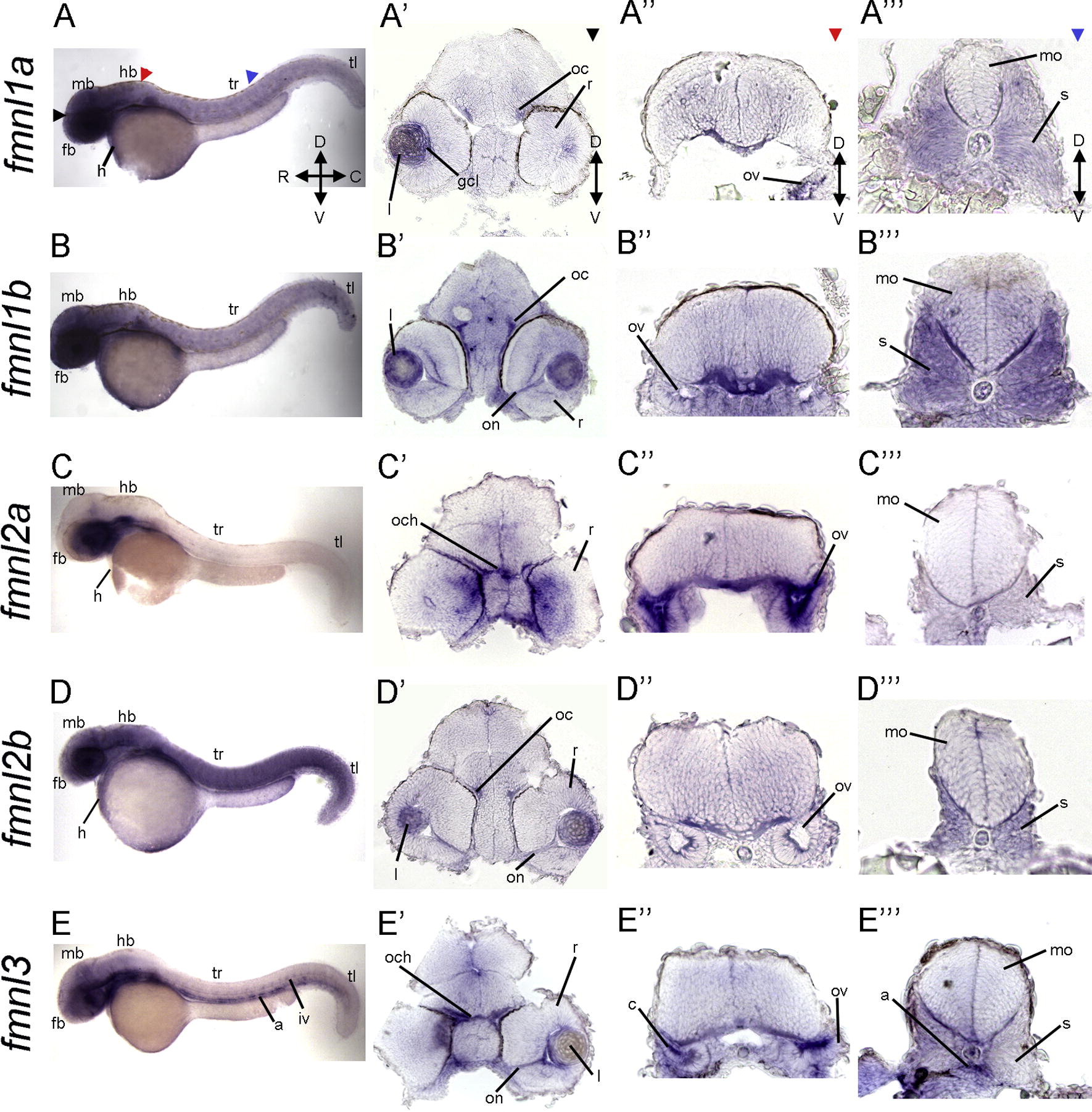
Fig. 6 Expression of fmnls at 48 hpf reveals more distinct patterns than at earlier stages. (A-E) Lateral views of whole-mount embryos. Colored triangles indicate level of cross sections: black indicates forebrain (fb; A?-E?), red at the level of the hindbrain (hb; A??-E??), and blue denotes sections through the trunk (tr; A????E???). Colored arrowheads indicate the level cross sections in (A?-E?) black arrows indicate forebrain (fb), (A???E??) at the level of the hindbrain (hb) red arrowhead and (A???-E???) blue arrowhead indicating sections through the trunk (tr). Enrichment of fmnl1a, 1b and 2b in the head (A, B, D). Robust expression of fmnl1a and 1b in somites (A??? and B???). fmnl1b, 2b and 3 expression in the visual system including the optic commissure (oc), optic nerve (on) and lens (l) (B?, D?, E?). fmnl2a expression in the medial region of the retina (C?). fmnl2a expression within the otic vesicle (ov), (C??), while (E??) Prominent expression of fmnl3 in the cristae (c). a, aorta; C, caudal; gcl, ganglion cell layer; D, dorsal; mb, midbrain; mo, medulla oblongata; n, notochord; och, optic chiasm; r, retina; iv, intersomitic vessels; R, rostral; r, retina; tl, tail; V, ventral.
Reprinted from Gene expression patterns : GEP, 13(1-2), Santos-Ledo, A., Jenny, A., and Marlow, F.L., Comparative gene expression analysis of the fmnl family of formins during zebrafish development and implications for tissue specific functions, 30-37, Copyright (2013) with permission from Elsevier. Full text @ Gene Expr. Patterns