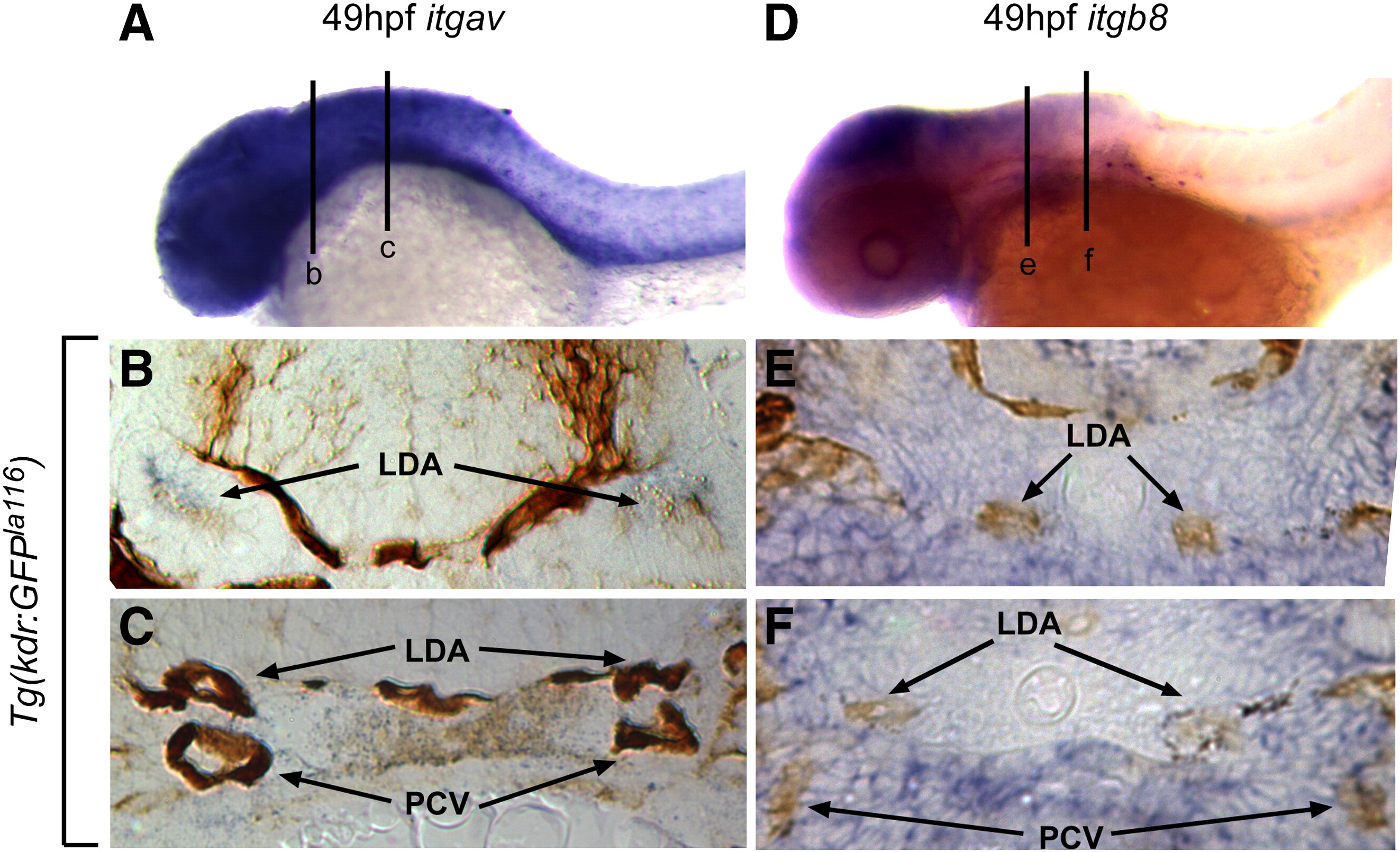
Fig. 2 Integrin αv and β8 are expressed in close proximity to head blood vessels. Comparative in situ hybridization of integrin αv (itgav; blue staining in A?C) or integrin β8 (itgb8; blue staining in D?F) as assayed in Tg(kdrl:eGFP)la116 embryos marking endothelial cells (brown staining in B, C, E, F). (A) At 49 hpf itgav shows broad expression in the head and anterior ventral trunk. (B) In a transverse section of the head (line b in A), itgav is expressed in close proximity to the lateral dorsal aortae (LDA). (C) A more posterior transverse section (line c in A) shows itgav expression in the ventral mesenchyme of the head (VM), surrounding the lateral dorsal aortae (LDA) and posterior cardinal veins (PCV). (D) Similarly, at 49 hpf, itgb8 is highly expressed in the head and weakly expressed in the anterior ventral trunk. (E) In a transverse section of the head (line e in D), itgb8 is expressed in the regions around the lateral dorsal aortae (LDA). (F). A more posterior transverse section (line f in D) shows itgb8 expression in the ventral mesenchyme of the head (VM), surrounding the lateral dorsal aortae (LDA) and posterior cardinal veins (PCV). LDA: lateral dorsal aorta; PCV: posterior cardinal veins.
Reprinted from Developmental Biology, 363(1), Liu, J., Zeng, L., Kennedy, R.M., Gruenig, N.M., and Childs, S.J., betaPix plays a dual role in cerebral vascular stability and angiogenesis, and interacts with integrin alphavbeta8, 95-105, Copyright (2012) with permission from Elsevier. Full text @ Dev. Biol.

