Fig. S1
Fig. S1
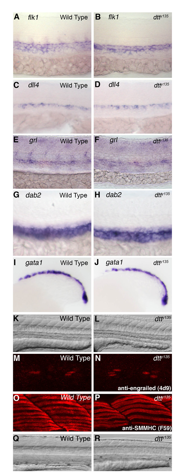
Endothelial, hematopoietic and muscle patterning is normal in dtty135 mutants. (A-J) In situ hybridization of the trunks of wild-type (A,C,E,G,I) and dtty135 mutant (B,D,F,H,J) zebrafish embryos probed for flk1 (A,B), dll4 (C,D), grl (E,F), dab2 (G,H) and gata1 (I,J). Marker expression appears equivalent in wild-type and dtty135 mutant animals. Embryos are at 26 hpf (A-H) and 18 hpf (I,J). (K,L) DIC transmitted light images of the trunks of 26 hpf wild-type (K) and dtty135 mutant (L) animals, showing normally patterned somites and muscles in dtty135 mutants. (M,N) Confocal imaging of trunks of wild-type (M) and dtty135 mutant (N) animals stained for immunofluorescence with a monoclonal anti-engrailed antibody (4D9), revealing normal muscle pioneer specification. (O,P) Confocal imaging of trunks of wild-type (O) and dtty135 mutant (P) animals stained for immunofluorescence with a monoclonal anti-SMMHC antibody (F59), revealing normal slow muscle differentiation. (Q,R) DIC transmitted light images of the trunks of 96 hpf wild-type (Q) and dtty135 mutant (R) animals, showing reduced muscle mass by 96 hpf in dtty135 mutants. Lateral views, anterior towards the left.

