
Fig. 3
|
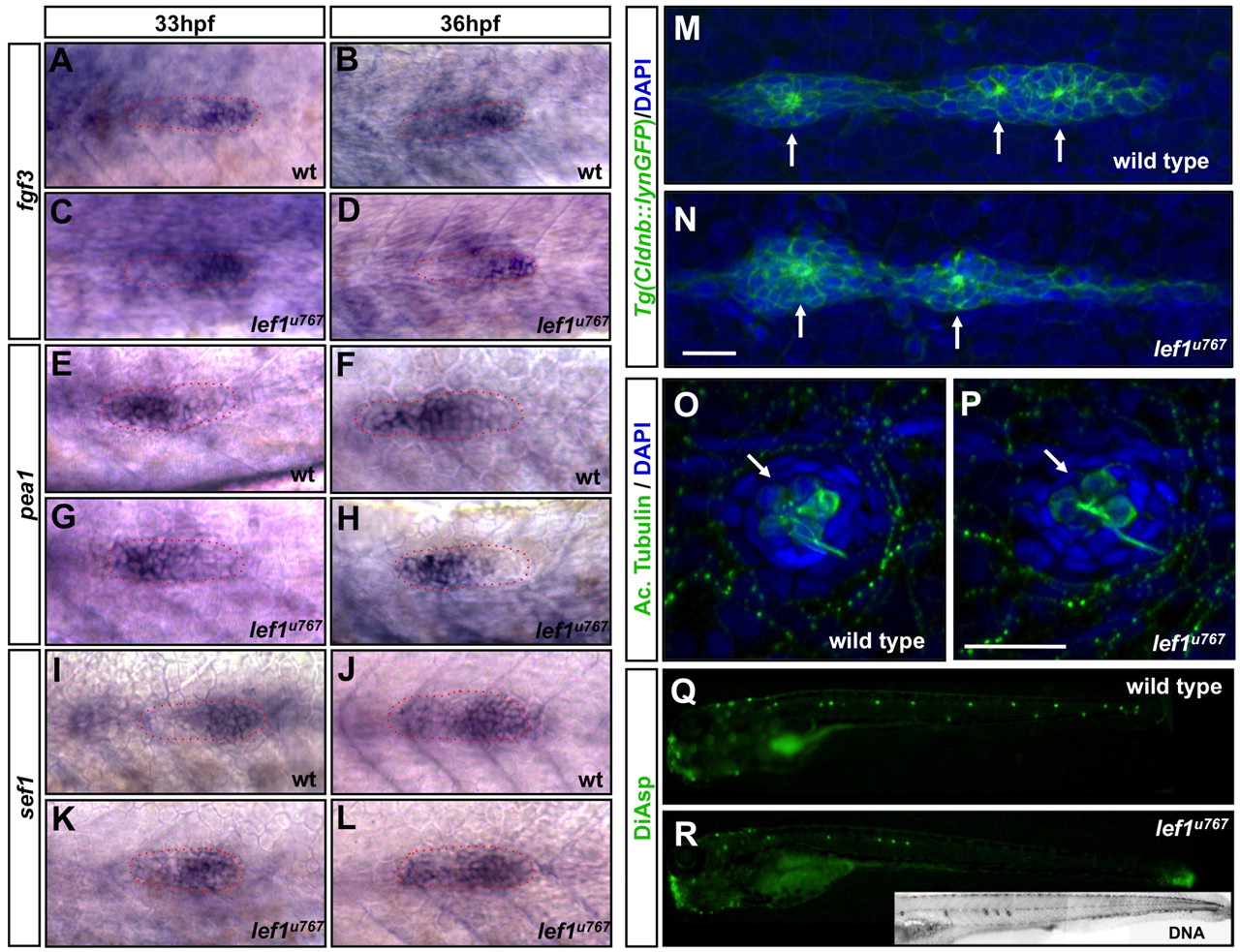
Fig. 3 Primordium organisation, neuromast morphology and differentiation are unaffected by loss of Lef1 function. (A-L) Expression of fgf3 (A-D), pea1 (E-H) and sef1 (I-L) in wild type (A,B,E,F,I,J) and lef1u767 mutants (C,D,G,H,K,L). Primordia are outlined. (M,N) Tg(-8.0cldnb:lynEGFP)zf106-labelled wild-type (M) and lef1u767 (N) primordia at 38 hpf. Membrane-anchored GFP reveals the polarisation of cells and their organisation into rosettes (arrows). (O,P) The last-deposited neuromast in a lef1u767 mutant (P) and wild type (O) labelled with anti-acetylated tubulin antibody (green). Nuclei are DAPI stained. Scale bar: 20 μm. (Q,R) Diasp staining in wild type (Q) and lef1u767mutant (R) at 3 dpf. DAPI-labelling of nuclei (inset in R) shows no more neuromasts in the lef1u767 other than those labelled with Diasp).