Fig. 1
Fig. 1
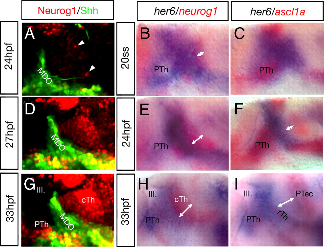
The neurogenetic gradient in fish. Glutamatergic neurogenesis spreads in a wave from posterior to anterior in the developing thalamus. Analysis of the dynamic expression of proneural genes during the development of the thalamic complex by in vivo imaging of double transgenic zebrafish and double in situ hybridisations. Upon induction of shh:GFP in the MDO, neurog1:RFP is induced first in the posterior Th (A, arrowheads). Over time, the caudal thalamus is filled with neurog1 positive cells (D and G). At the 20-somite stage, neurog1 mRNA can be detected within the thalamic complex (B). Over time, the expression increases from posterior to anterior and fills the entire cTh at 33 hpf (E and H). At 20 somite stage, her6 expression marks the entire thalamic complex (B and C) and is gradually down-regulated in the caudal part over time. her6 expression is maintained in the PTh, MDO, rTh and absent from the cTh at 33 hpf (H and I). The expression domains of her6 and neurog1 are abutting. In contrast to neurog1, the proneural gene ascl1a is induced from ventral to dorsal within the her6 positive PTh and rTh from the 20-somite stage (C), to 24 hpf (F) and 36 hpf (I). Embryos are shown laterally, white double-headed arrows indicate the increasing width of the cTh. III, third brain ventricle; cTh, caudal Thalamus; MDO, mid-diencephalic organizer; PTh, prethalamus; rTh, rostral Thalamus; ss, somite stage.

