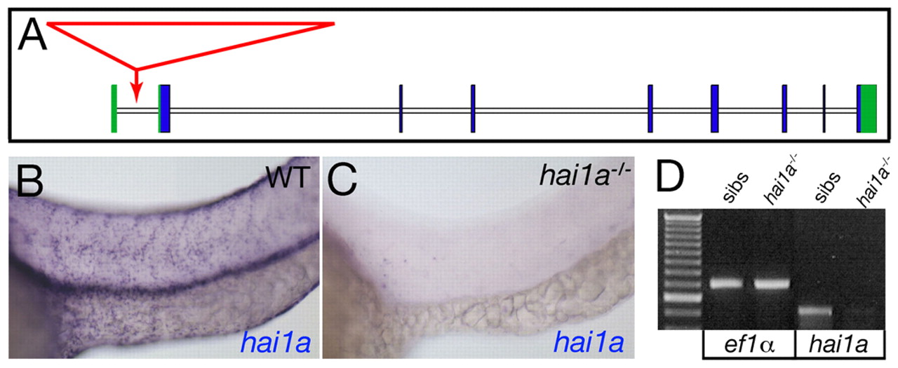
Fig. 2 The insertion in hi2217 abrogates hai1a transcription. (A) Structure of the hai1a gene showing the viral insertion site in the hi2217 allele (red). Exons are boxed with coding and non-coding sequences in blue and green, respectively. The viral insertion (red arrow) occurs in the first intron upstream of the first coding exon. (B,C) Lateral views of the trunk epidermis of a wild-type sibling (WT; B) and a hi2217 homozygote (C) at 24 hpf, after hai1a in situ hybridization. (D) Reverse transcriptase (RT)-PCR analysis of hi2217 homozygotes (lanes 3,5) and wild-type siblings (lanes 2,4) at 24 hpf, demonstrating a strong reduction in hai1a transcript levels in mutants compared with siblings, whereas ef1a levels are identical. Lane 1, 100 bp ladder.
Image
Figure Caption
Figure Data
Acknowledgments
This image is the copyrighted work of the attributed author or publisher, and
ZFIN has permission only to display this image to its users.
Additional permissions should be obtained from the applicable author or publisher of the image.
Full text @ Development

