Fig. 5
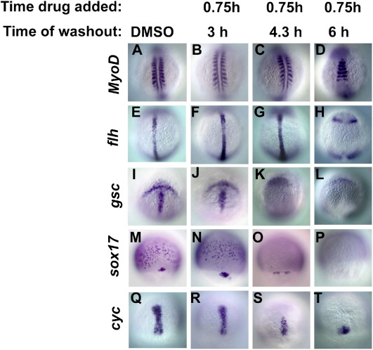
Fig. 5 Progressive loss of cell fates with shorter exposures to Activin-like signals. Embryos were treated with DMSO (A, E, I, M, Q) or with SB-505124 between 0.75 hpf and 3 hpf (B, F, J, N, R), 4.3 hpf (C, G, K, O, S) or 6 hpf (D, H, L, P, T). Sibling embryos were divided into five groups. One group was fixed at 14 hpf and stained for MyoD expression (A?D). Embryos fixed at 10 hpf were stained for either flh (E?H), or gsc (I?L). Embryos fixed at 8 hpf were stained for either sox17 (M?P) or cyc (Q?T). When the drug is washed out at 3 hpf (B, F, J, N, R), expression of all markers is indistinguishable from wild type (A, E, I, M, Q). When the drug is washed out at 4.3 hpf, MyoD and flh expression is normal (C, G), but gsc is absent from the prechordal plate (K) and sox17 is only expressed in the dorsal forerunner cells (O). The axial domain of cyc expression is reduced (S). When the drug is washed out at 6 hpf, MyoD is expressed in trunk somites (D), and flh is expressed in four ectodermal domains, but not in the axial mesoderm (H). gsc is absent from the prechordal plate (L) and sox17 expression is abolished (P). cyc expression in the axial mesoderm is severely reduced, but is still present (T). In panels A?L, images are dorsal views, with anterior to the top of the page. In panels M?I, images are dorsal views with the animal pole at the top.
Reprinted from Developmental Biology, 309(2), Hagos, E.G., Fan, X., and Dougan, S.T., The role of maternal Activin-like signals in zebrafish embryos, 245-258, Copyright (2007) with permission from Elsevier. Full text @ Dev. Biol.

