Fig. 2
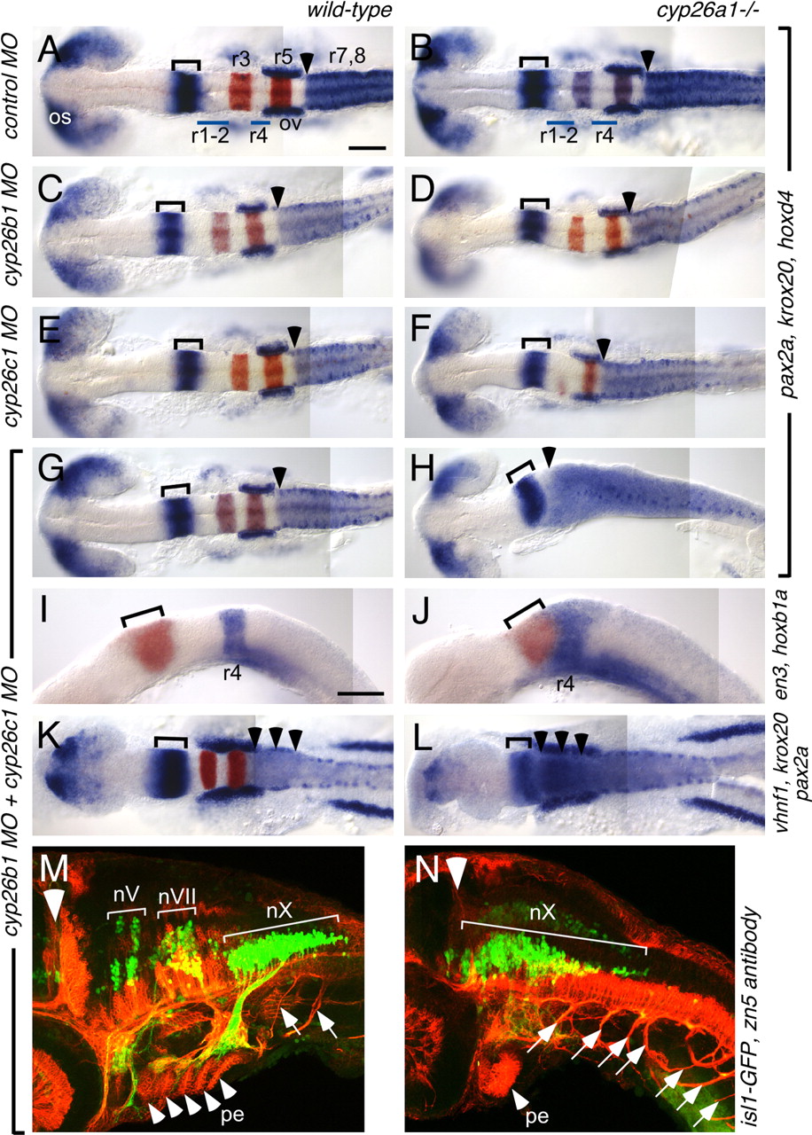
Fig. 2 cyp26b1 and cyp26c1 function redundantly with cyp26a1 to pattern the hindbrain. Whole-mount RNA in situ hybridizations at 18 hpf (A-J) and 13 hpf (K,L) and immunostaining at 48 hpf (M,N) in wild-type (left column) and cyp26a1-/- (right column) embryos injected with MOs as shown on the left. (A-H) pax2a (blue) marks the optic stalk (os), posterior midbrain and cerebellum (bracket), and the otic vesicles (ov); whereas hoxd4 (also blue) marks the r7-r8 territory and krox20 (red) marks r3 and r5. MO depletion of Cyp26b1 and/or Cyp26c1 does not affect this pattern in wild-type embryos (C,E,G), but progressively posteriorizes the hindbrain in cyp26a1-/- embryos (D,F,H). Arrowhead marks the r6-r7 boundary, which is shifted to the anterior hindbrain in Cyp26-depleted embryos. (I,J) en3 (red) marks the posterior midbrain and cerebellum (bracket) and hoxb1a (blue) marks r4, which is shifted anteriorly in Cyp26-depleted embryos. (K,L) pax2a (blue) and krox20 (red) are expressed as described above. vhnf1 (also blue) is expressed in the posterior hindbrain up to the r5-r6 boundary (arrowheads) and is also shifted anteriorly in Cyp26-depleted embryos. (M,N) The isl1-GFP transgene (green) marks cranial motor neurons (nV: trigeminal motor neurons in r2 and r3; nVII: facial motor neurons in r4-6; nX: vagal motor neurons in r8) whereas the zn5 antibody (red) marks spinal motor neurons (arrows), pharyngeal arch endoderm (pe, arrowheads mark individual pharyngeal arches) and other structures. The large white arrowhead indicates the mid-hindbrain boundary. In Cyp26-depleted embryos, the motor neurons of the vagus nerve (nX) are expanded anteriorly, as are the spinal motor neurons. Scale bars: 100 μm. Scale bar in A is for A-H,K,L; scale bar in I is for I,J.

