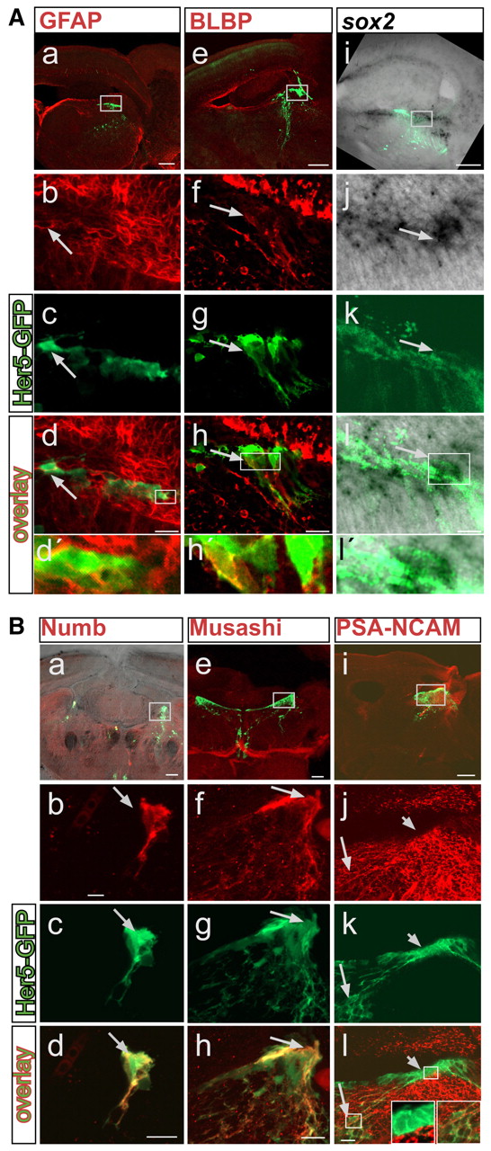
Fig. 4 Her5-GFP-positive cells lining the tectal ventricle express stem cell markers. (A) Co-localization of Her5-GFP (green) with GFAP (a-d'), BLBP (e-h'), sox2 (i-l'). (B) Co-localization of Her5-GFP (green) with Numb (a-d), Musashi (e-h) and PSA-NCAM (i-l). Sagittal (GFAP, BLBP, sox2, PSA-NCAM) or cross-sections (Numb and Musashi) are shown. sox2 is detected by in situ hybridization (black signal), while all other markers are detected by immunohistochemistry (red signal). (A, parts i-l'; B, part a) An overlay with the brightfield view in addition to fluorescence. All long arrows point to cells that co-express Her5-GFP and the respective markers. (B, parts j-l) Her5-GFP-positive cells located close to the ventricle, which also express her5 RNA (Fig. 1C,E), are PSA-NCAM-negative (short arrow, left inset) while Her5-GFP-positive cells located further ventrally do express PSA-NCAM (long arrow, right inset), indicating their differentiation into a neuronal fate. Scale bars: 100 μm in a,e,i; 10 μm in b-d,f-h,j-l.
Image
Figure Caption
Figure Data
Acknowledgments
This image is the copyrighted work of the attributed author or publisher, and
ZFIN has permission only to display this image to its users.
Additional permissions should be obtained from the applicable author or publisher of the image.
Full text @ Development

