
Fig. 6
|
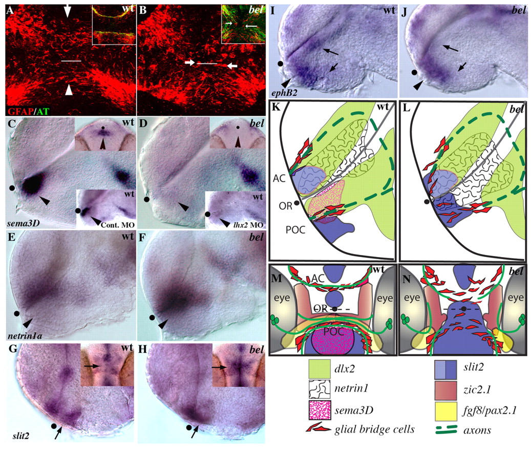
Fig. 6 The axon growth substrate is disrupted in bel mutants. (A, B) Confocal images of ventral views of 21 hpf embryos labeled with anti-GFAP antibody (red); white line marks the optic recess. (A) GFAP expressing glial cells span the midline in the position of the AC (arrow) and POC (arrowhead). (B) In bel mutants, these glial bridges are disorganized and glial cell bodies are present between the commissures (arrows). Insets show a similar disorganization of glial cells in the mutant at 28 hpf in embryos double labeled for axons (anti-AT, green) and glial cells (anti-GFAP, red). (C, D) sema3d is absent in the forebrain of the bel mutants (arrowhead). Upper insets show ventral views; lower insets show a similar loss of sema3d expression in the anterior diencephalon after injection of lhx2 antisense MO into a wild-type embryo. (E, F) netrin1a expression is expanded ventrally into the diencephalon in bel mutants (arrowhead). (G, H) Expression of slit2 in bel mutants is expanded across the chiasm region (arrows). Insets show ventral views, anterior up. (I, J) ephB2 expression is reduced in the diencephalon and hypothalamus (arrows), but appears normal in the chiasm region (arrowheads). (K-N) Schematic lateral (K,L) and ventral (M,N) views showing expression of a subset of the genes described in this and the previous figure. AC, anterior commissure; OR, optic recess; POC, post optic commissure; black dots mark the position of the optic recess.