|
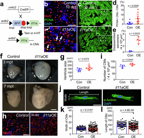
Myocardial il11a overexpression (OE) triggers CM proliferation in the absence of injury. a Schematic of tamoxifen (Tam) or 4-HT (4-Hydroxytamoxifen) inducible il11a overexpression (OE). Tg(actb2:loxp-TagBFP-STOP-Loxp-il11a); cmlc2:CreER were administered with Tam or 4-HT to overexpress il11a (il11aOE). CreER-negative littermates were used as controls. b Representative cardiac section images stained with Mef2 (Red) and PCNA (Green) from control and il11aOE at 10 days post-treatment (dpt). Arrows indicate proliferative CMs. c Representative cardiac sections images stained with α-Actinin (green) from control and il11aOE uninjured hearts at 30 dpt. Insets correspond to higher magnifications of dashed boxes. d The percentage of PCNA+ CMs of uninjured hearts from control (Con) and il11aOE (OE) at 7 dpt. Biological replicates = 11 for control and il11aOE. e Quantification of disorganized sarcomere structures in the ventricles of Con and il11aOE at 30 dpt. Biological replicates = 3 and 4 for control and il11aOE, respectively. f Whole-mount images of 3- and 7-months post treatment (mpt) hearts from Con and il11aOE. g Quantification of the ventricle size from 3 mpt Con and il11aOE. Biological replicates = 8 for control and il11aOE. h Representative cardiac section images of 30 dpt control and il11aOE stained with Mef2 (red) and DAPI (blue). i Quantification for CM numbers in the ventricles from 30 dpt Con and il11aOE. Biological replicates = 5 for control and il11aOE. j Representative images of dissociated ventricular CMs stained with α-Actinin (green) from 2 mpt control and il11aOE. Quantification of the width (k) and the length (l) of individual CMs. Biological replicates = 92 and 234 for control and il11aOE, respectively. Scale bars, 50 µm in (b), (c) and (h), 0.5 mm in (f), 25 µm in (j). Data are mean ± SEM in (d), (e), (g), (i), (k), and (l). p-values were determined by unpaired two-tailed t-test in (d), (e), (g), (i), (k), and (l).
|