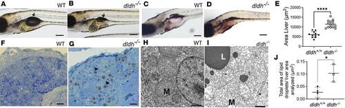
Liver pathology in dldh?/? zebrafish larvae. (A and B) WT and dldh?/? morphology at 7 dpf showed mutants had increased liver size (yellow line) and deflated swim bladder (arrow). Scale bar: 100 ?m (�2.5 magnification of the middle fish in Figure 4B). (C and D) Oil Red O staining of 8 dpf WT and dldh?/? liver showed increased lipid accumulation in mutant larvae. Scale bar: 100 ?m. (E) Liver area (?m2) was increased in 14 dldh?/? relative to 11 WT larvae by ImageJ analysis across 3 replicate experiments. ****P < 0.0001 by unpaired Student?s t test. (F and G) Thin sections of WT and dldh?/? liver stained with toluidine blue showed mutant larvae had increased frequency of lipid droplets (arrowheads). Scale bars: 10 �m. (H and I) Hepatocyte ultrastructure in 7 dpf WT and dldh?/? showed mutant larvae had mitochondrial damage (M) and large lipid droplets (L). Scale bars: 1 �m. (J) Ultrastructural analysis showed increased fractional lipid droplet to liver area analyzed in 4 WT and 3 dldh?/? larvae at 7 dpf. Areas determined using ImageJ. *P < 0.05 by unpaired Student?s t test.
|
