
Fig. 6
- ID
- ZDB-FIG-240904-17
- Publication
- Sam et al., 2024 - Gata6 functions in zebrafish endoderm to regulate late differentiating arterial pole cardiogenesis
- Other Figures
- All Figure Page
- Back to All Figure Page
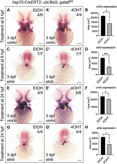
Gata6 regulates the addition of late differentiating cardiomyocytes. (A-D) Hsp70:CreERT2; ubb:BsG; gata6fl/fl embryos phenocopy gata6wcm7/wcm7 cardiac defects. Whole-mount in situ hybridization (WISH) analysis revealed decreased expression domains of both vmhc (A,A?) and elnb (C,C?) transcripts in gata6fl/fl embryos after tamoxifen treatment at 6 hpf. (B,D) Quantification of the area of WISH staining showed a statistically significant loss of the mRNA expression domain within the ventricle (B) and outflow tract (D) (paired t-test, *P<0.05 and ****P<0.0001). Data are mean�s.e.m. (E-H) Relatively late removal of gata6 by heat-shock at 24 hpf still results in reduced cardiac tissue. WISH staining showed decreased expression domains of vmhc (E,E?) and elnb transcripts (G,G?) in gata6fl/fl embryos after tamoxifen treatment at 24 hpf. (F,H) Quantification of the area of WISH staining in the ventricle (F) and outflow tract (H) (paired t-test, *P<0.05). Data are mean�s.e.m. Scale bars: 100 �m. White brackets indicate the length of the ventricle in ethanol-treated controls. Yellow bars indicate the length of the ventricle in tamoxifen-treated embryos. Black arrows indicate the reduced area of elnb expression. Area of staining was measured manually using FIJI software. |