
Fig. 3
- ID
- ZDB-FIG-240904-14
- Publication
- Sam et al., 2024 - Gata6 functions in zebrafish endoderm to regulate late differentiating arterial pole cardiogenesis
- Other Figures
- All Figure Page
- Back to All Figure Page
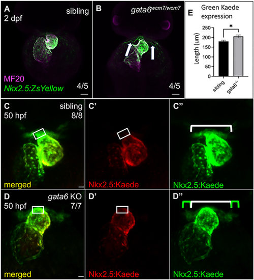
Loss of gata6 impairs the addition of Nkx2.5+ progenitors to the arterial pole. (A,B) Immunostaining at 2 dpf (frontal view) shows Nkx2.5:ZsYellow+ progenitors within and surrounding the heart in both siblings and mutants. Excess ZsYellow+ cells remain proximal to the ventricle in the mutants (white arrows). Scale bars: 50 �m. (C-D?) Representative confocal images of 50 hpf Tg(nkx2.5:kaede) embryos after photoconversion of the heart tube at 25 hpf. Tg(nkx2.5:kaede); gata6?/? embryos express more unconverted Kaede protein (D?, green bar) around the arterial pole compared with wild type or heterozygous siblings (C?, white bar, also shown in D? for comparison). White rectangles indicate the region of cardiac cells at the arterial pole that are green+/red? (late differentiating) in sibling hearts and green+/red+ (early differentiating) in mutant hearts. Scale bars: 20 ?m. (E) Quantification of the green Kaede region expressed outside the heart as marked in C? and D? showing significantly more cardiac cells accumulated around (but not on) the ventricle in mutants compared with siblings (paired t-test, *P<0.05, data are mean�s.e.m.). Length was measured manually using FIJI software. |