
FIGURE
Fig. 4
Fig. 4
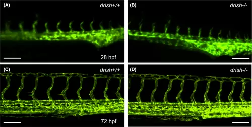
drish mutants do not show defects in vascular patterning. (A?D) Confocal images of Tg(kdrl:GFP) expression in drish+/+ and drish?/? mutant embryos at 28 and 72 hpf using �20 and �10 objectives. drish?/? mutants exhibit normal vascular patterning (B, D), similar to drish+/+ embryos (A, C). Scale bars: 100 ?m. |
Expression Data
Expression Detail
Antibody Labeling
Phenotype Data
Phenotype Detail
Acknowledgments
This image is the copyrighted work of the attributed author or publisher, and
ZFIN has permission only to display this image to its users.
Additional permissions should be obtained from the applicable author or publisher of the image.
Full text @ Dev. Dyn.