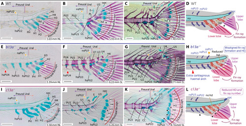
Ontogenetic series of cleared and stained skeletal preparations for b13a?/? and c13a?/? mutants. The figure displays the development of the caudal fin in zebrafish WT, b13a?/?, and c13a?/? mutants, focusing on the formation of fin rays and posterior vertebrae. (A) 5.03-mm NL WT specimen. (B) 5.93-mm SL WT specimen. (C) 7.40-mm SL WT specimen. (D) Diagram showing normal zebrafish caudal fin development, fin ray appearance is aligned with the hypural diastema. (E) 5.38-mm NL b13a?/? mutant specimen. (F) 6.22-mm SL b13a?/? mutant specimen. (G) 8.71-mm SL b13a?/? mutant specimen. (H) Diagram showing the b13a?/? mutant caudal fin development, with a reduced hypural diastema and the appearance of fin rays not aligned with the position of the diastema. The notochord is abbreviated, with no opisthural cartilage, the base of hypurals is closer to the notochord posterior end, and an additional preural element is present anteriorly. (I) 5.58-mm NL c13a?/? mutant specimen. (J) 6.26-mm SL c13a?/? mutant specimen. (K) 8.16-mm SL c13a?/? mutant specimen. Black arrowheads, extra distal radials. (L) Diagram showing the c13a?/? mutant caudal fin development, lacking a hypural diastema, with fewer fin rays forming, more spaced between them, and additional radials at the distal ends of the hypurals 1 and 2. Red arrowheads, extra cartilaginous haemal arch or spine; yellow arrowheads, first neural arch/spine that develops; black asterisk, separation between upper and lower caudal fin lobes. See Fig. 1 for anatomical terminology. Scale bars, 0.1 mm.
|
