|
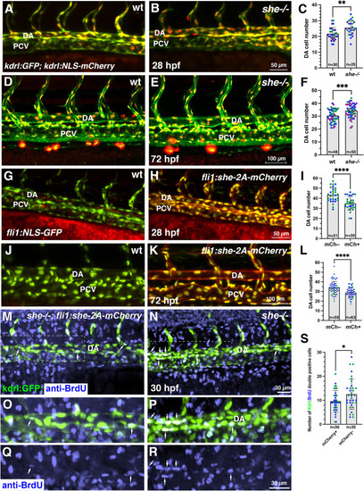
she affects cell number and inhibits endothelial cell proliferation. (A-F) Analysis of cell number in the DA of she mutant and wild-type (she+/+) sibling embryos at 28 and 72 hpf. Embryos were obtained from an incross of she+/-; fli1:GFP; kdrl:NLS-mCherry parents, imaged by confocal microscopy and subsequently genotyped. Note increased cell number in she mutant embryos. (G-L) Analysis of cell number in the DA of fli1:she-2A-mCherry; fli1:NLS-GFP embryos and their mCherry negative (wt) siblings at 28 and 72 hpf. Note the reduced cell number in mCherry+ embryos. (M-S) Cell proliferation analysis using BrdU incorporation assay in she-/-; fli1:she-2A-mCherry embryos (phenotypically normal) and their sibling mCherry negative she-/- embryos in kdrl:GFP background at 30 hpf. (O-R) show enlarged view of the area in insets (M,N). Note the increased number of BrdU and kdrl:GFP double positive cells within the DA in mCherry-negative she mutant embryos. All graphs show data combined from 2 (I,S) or 3 (C,F,L) independent experiments; data points from different experiments are shown in distinct colors. Mean±SD is shown. *p<0.05, **p<0.01, ***p<0.001, ****p<0.0001, Student’s t-test.
|