|
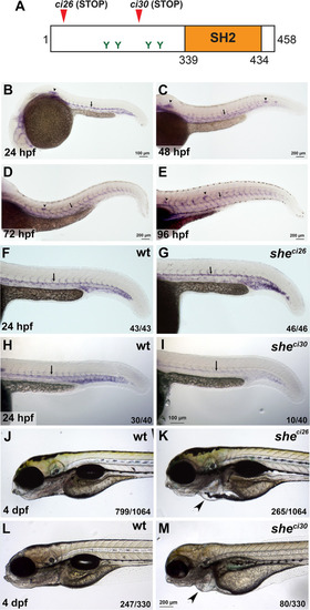
she mutants display pericardial edema and loss of blood circulation. (A) Zebrafish SHE protein diagram. she ci26 and ci30 mutant alleles are predicted to result in a frameshift and premature stop codons. SH2 domain and consensus tyrosine ABL phosphorylation sites are shown. (B-E) In situ hybridization analysis of she expression in wild-type embryos at 24, 48, 72 and 96 hpf stages. Note its expression in the dorsal aorta (arrows), intersegmental vessels, lateral line primordium (arrowhead, B) and neuromasts (arrowheads, C-E). (F-I) In situ hybridization analysis of she expression in she mutants at 24 hpf. Note that she expression is unaffected in sheci26 embryos while it is strongly reduced in sheci30 embryos. Homozygous sheci26 mutant embryos were obtained by an incross of sheci26-/-; fli1a:she-2A-mCherry; kdrl:GFP parents (which are viable due to fli1a:she-2A-mCherry rescue) and selected for mCherry negative embryos. Wild-type control embryos were obtained by incross of sibling wt; fli1a:she-2A-mCherry parents and selected for mCherry-negative embryos. sheci30 embryos were obtained by incross of sheci30+/- parents and genotyped after in situ hybridization. 25% (10 out of 40) embryos showed strong reduction in she expression which correlated with the mutant phenotype. (J-M) Brightfield images of sheci26 and sheci30 mutant embryos and their siblings (wild-type and or heterozygous) at 4 dpf. Note the pericardial edema (arrowheads) in the mutant embryos. Embryos were obtained by the incross of heterozygous parents in kdrl:GFP background. 24.9% (265 out of 1064) and 24.2% (80 out of 330) embryos obtained in sheci26 or sheci30 incross, respectively, showed this phenotype. A subset of embryos was genotyped to confirm the correlation between the phenotype and genotype.
|

