
Fig. 6
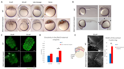
View largeDownload slide Fhdc3 is necessary to maintain the yolk-blastodisc boundary during early development. (A) Images of live embryos comparing fhdc3 maternal crispant embryos with time-matched wild-type (WT) controls during embryonic development. An abnormal constriction at the boundary between the blastodisc and yolk (black arrowhead) is observed during the early cleavage stages (64-cell). At later stages, a normal degree of constriction at the yolk-blastodisc boundary is restored, but embryos exhibit presumptive ectopic yolk inclusions (blue arrows). (B) At 1 dpf, fhdc3 maternal crispant embryos do not exhibit gross morphological defects, although they still contain presumptive ectopic yolk inclusions (arrows). (C) Immunolabeling of β-catenin and DAPI staining at 8-cell and 64-cell stages, showing that the overall shape and cell organization of the embryo are affected in the later fhdc3 maternal crispant embryos. (D) At the 8-cell stage, there is no significant difference in the shape of wild-type and fhdc3 maternal crispant embryos [fhdc3 maternal crispant score of circularity 0.881 (n=18); wild type 0.8615 (n=13); P=0.0893] but the shape appears significantly different at the 64-cell stage [fhdc3 maternal crispants: 0.90963 (n=8), wild type: 0.84757 (n=7); P=0.0003]. Error bars represent s.e.m. (E) At the 16-cell stage, the cortical F-actin ring appears significantly wider in the fhdc3 maternal crispant [fhdc3 maternal crispants: 24.8 μm (n=10), wild type: 14.1 μm (n=10); P=0.0003]. Error bars represent s.e.m. Brackets indicate the width of the cortical F-actin ring at the location shown in the schematic. Scale bars: 100 μm (C); 20 μm (E). |