
Fig. 4
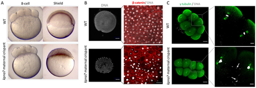
Kpna7 is necessary for nuclear segregation during early development. (A) Images of live kpna7 maternal crispant and wild-type (WT) controls at the 8-cell stage (75 mpf) and shield stage (6 hpf). At the 8-cell stage, kpna7 maternal crispants appear to divide normally, but they stall at the sphere stage and fail to undergo epiboly. (B) Immunohistochemistry labeling of β-catenin and DAPI staining at 6 hpf showing that the kpna7 maternal crispant embryos exhibit nuclei of unequal sizes, including a subset of cells that entirely lack nuclei (blue asterisks). (C) Immunohistochemistry labeling of γ-tubulin and DAPI staining at 75 mpf, showing that kpna7 maternal crispant embryos display abnormal nuclear segregation (arrows) during cell cleavage. Scale bars: 100 μm (B,C, low magnification); 20 μm (B,C, high magnification). |