FIGURE

Figure 4

ID
ZDB-FIG-231016-74
Publication
Hodges et al., 2023 - Disrupting the Repeat Domain of Premelanosome Protein (PMEL) Produces Dysamyloidosis and Dystrophic Ocular Pigment Reflective of Pigmentary Glaucoma
Other Figures
All Figure Page
Back to All Figure Page
Figure 4

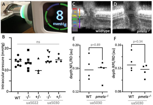
Adult pmela−/− mutants presented no measurable phenotypes when evaluated via rebound tonometry and ocular computed tomography (OCT). (A,B) No consistent difference in intraocular pressure (IOP) was apparent among adult zebrafish of various pmela genotypes. IOP was measured in adult zebrafish using Tonovet plus. Average readings for each fish are plotted. A Kruskal–Wallis statistical test showed no significant differences between means. (CF) Retinal lamination and layer depths showed no overt alterations when pmela mutants were assessed in vivo via OCT. Exemplar images are shown for adult wildtype and ua5030 homozygotes, whereas ua5022 homozygotes and measurements of additional layers are reported in Figures S3 and S4. The ratio of each retinal layer was normalized to the full retinal depth (RD) for each fish. RPE, retinal pigment epithelium; PL, photoreceptor layer; ONL, outer nuclear layer; INL, inner nuclear layer; IPL, inner plexiform layer; NFL, neurofibrillary layer. Comparison with age-matched controls used Mann–Whitney statistical tests. The horizontal lines in the graphs B, E and F represent the mean values of multiple fish and the value for each individual fish is plotted as a filled symbol (circle, square, rectangle, diamond, etc.).

Expression Data

Expression Detail
Antibody Labeling
Phenotype Data
Fish:
Observed In:
Stage: Adult

Phenotype Detail
Acknowledgments
This image is the copyrighted work of the attributed author or publisher, and ZFIN has permission only to display this image to its users. Additional permissions should be obtained from the applicable author or publisher of the image. Full text @ Int. J. Mol. Sci.